
What does the treatment look like and how easy is it for the patient?
See how easily the patient carries out the daily adjustment at home. The compact TrueLok™ Elevate unilateral fixator sits on the shin, keeping the foot 100% free for dressings, offloading, and daily life with close medical supervision.
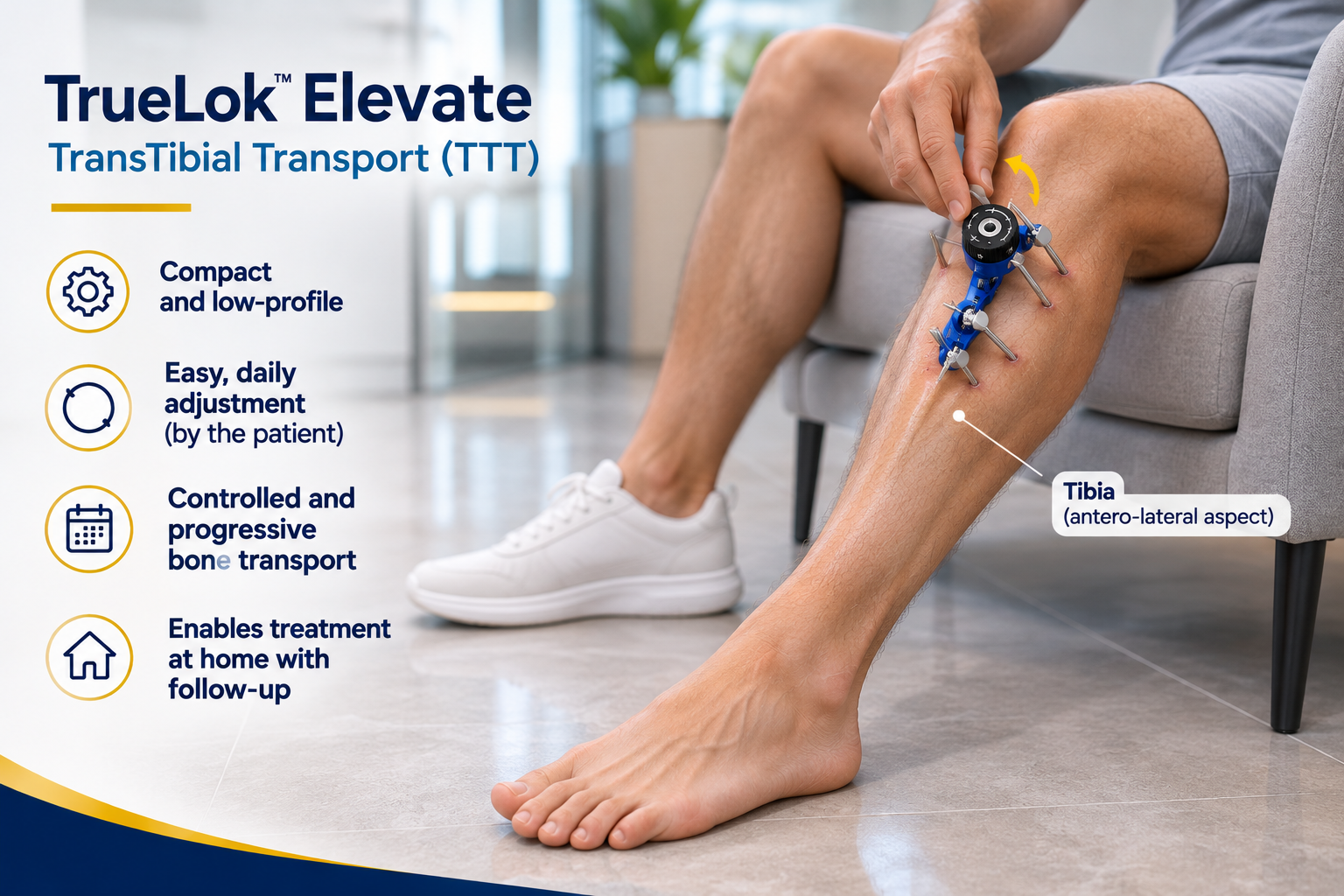
Compact & low-profile
Lightweight unilateral fixator on the shin without bulky full-ring frames.
Easy daily adjustment
The patient simply turns the dial gently 1 mm per day according to protocol.
Progressive transport
Controlled mechanotransduction that stimulates new capillary vessels to the foot.
Home recovery
Carried out at home with regular outpatient clinic checkups and foot wound care.
When is it worth assessing a foot lesion?
Not every callus or scrape needs a specialist. But certain situations deserve a focused assessment to understand what is slowing healing and what options may help. New or persistent lesions need clinical assessment — and some symptoms need urgent care.
A wound that won't heal
A lesion on the foot, toes or heel that has not closed in the expected time despite care.
An active ulcer
An open ulcer on the foot, toes or heel, especially in someone with diabetes.
A lesion that keeps coming back
A wound that reappears after healing, suggesting persisting risk factors underneath.
Colour or temperature changes
Redness, dark discolouration, or a foot that feels cooler or warmer than expected.
Loss of sensation
New numbness, tingling or reduced feeling in the feet — a common diabetic change.
Pain when walking
Leg or calf symptoms on exertion that ease with rest, which can relate to circulation.
Previous ulcer or amputation
A history of foot lesions or toe amputation raises the risk of new problems.
Diabetes with reduced circulation
Diabetes combined with vascular disease significantly increases foot-wound risk.
An assessment of the factors that affect how a wound heals
Every wound is different. A specialist assessment looks at the combination of factors that may be holding back healing — from blood supply and sensation to pressure, infection and overall health.
A. Blood supply and perfusion
How well the foot and leg are being reached by blood flow — a key factor in whether a wound can heal.
B. Sensation and neuropathy
Whether loss of protective sensation is present, which can allow injuries to go unnoticed.
C. Infection and wound size
Signs of infection and the surface extent of the lesion, which influence how it is managed.
D. Depth and tissues involved
How deep the ulcer reaches and which structures — skin, tendon, bone — are affected.
E. Pressure, deformity and offloading
Pressure points under the foot, deformities, and the need to reduce load on the wound.
F. Blood sugar and related conditions
Metabolic control and other medical conditions that can slow healing.
G. History and previous treatments
Your clinical background and what has been tried so far, to understand what has and hasn't helped.
H. Further tests or other specialties
Whether additional studies or input from another speciality (for example vascular) are needed.
Not every test is carried out at the clinic. The assessment determines what is indicated for you and where it should be done.
Four areas of specialist support
Active ulcer assessment
- Identifying the factors slowing your wound's healing
- Considering debridement and local wound care where indicated
- Assessing infection and the need for additional attention
- Offloading pressure from the wound when appropriate
- Vascular review when arterial disease is suspected
- Following the clinical evolution over time
What is indicated is decided individually — not every measure is needed for every wound, and availability is confirmed during your assessment.
Preventing recurrence after healing
- Reassessment once the lesion has closed
- Identifying persisting risk factors
- Individualised footwear and offloading advice
- Monitoring to catch new lesions early
- Education to recognise warning signs
Prevention is tailored to your risk profile; no measure guarantees a lesion will never return.
High-risk diabetic foot prevention
- Assessment of sensation and circulation where indicated
- Identifying deformities and high-pressure zones
- Individual risk stratification
- Preventive recommendations
- Coordination with other care when needed
A preventive assessment can help, especially before an ulcer appears, but it does not eliminate risk entirely.
Circulation: PAD and claudication
- Assessment of circulation-related symptoms
- Review of their functional impact on walking
- Reviewing available tests and prior results
- Guidance on medical management and structured exercise where appropriate
- Vascular review or referral for revascularisation when indicated
Management is individualised — walking does not improve every form of arterial disease, and a single technique does not resolve all circulation symptoms.
Individualised options for complex lesions
Often a combination of measures
Treating a complex ulcer may combine established wound care, offloading, infection management, vascular review and addressing the underlying diabetes and circulation. The right mix depends on your individual assessment.
Established, complementary and limited-evidence options
Some measures are well established, some are complementary options for selected cases, and others still have limited or investigational evidence. We distinguish these clearly and do not present an option as a guaranteed result.
Limb preservation is a goal, not a guarantee
Where advanced or surgical options are indicated, the assessment helps clarify which may be appropriate for you, together with their limitations, risks and alternatives. The procedures currently available as part of the service are confirmed during your assessment and on enquiry — we do not list techniques that are not confirmed as part of the service.
Documented clinical cases
Authorised, documented cases will appear here
We do not publish invented patients, fabricated before-and-after photographs, or unverified results. When documented clinical cases — with the patient's consent and in line with privacy and advertising rules — are available, they will be shown here with the relevant clinical context and limitations.
A specialist assessment adapted to each patient
Led by Dr. Pablo Banchero
Orthopaedic surgeon Dr. Pablo Banchero leads the Traumatology Team clinical pathway. He speaks English, Spanish and Italian and assesses the orthopaedic and surgical aspects of complex foot and lower-limb lesions.
Read about Dr. BancheroA coordinated specialist network
Vascular and other specialist areas are covered through the Traumatology Team specialist network, with verified specialists joining as the network grows. We do not name professionals who are not yet confirmed. Where your case needs another speciality, we help coordinate that input.
Meet the teamHow your first assessment works
Initial contact
Reach out by WhatsApp, phone or our enquiry form to tell us what is troubling you.
Case review
Relevant information is gathered and the best way to organise your assessment is decided.
Specialist assessment
The lesion, your history, risk factors and available tests are reviewed.
Individualised plan
Appropriate options, follow-up needs and any referral to other specialties are explained.
Not everyone receives a specific procedure at the first visit, and we do not promise healing timelines or guaranteed outcomes. The aim of the first assessment is to understand your situation and explain what is indicated for you.
Transverse Tibial Bone Transport (TTT)
Transverse tibial bone transport (TTT) is a surgical technique that mobilises a controlled segment of the tibial cortex using a bone fixation and transport device. It has been studied primarily as a complementary treatment in selected patients with diabetic foot ulcers and peripheral microcirculation impairment, with the aim of inducing biological and vascular responses that may help improve tissue perfusion and support healing.
How does the external fixator look and where is it placed?
The compact unilateral fixator is mounted on the medial aspect of the proximal tibia (shin). It does not touch the foot, allowing daily dressings, debridement and ulcer offloading to continue freely.
1. Controlled osteotomy
Superficial cut in proximal tibial cortex preserving leg alignment and load-bearing stability.
2. Bone block
Cortical bone block slowly translated millimeter by millimeter toward the device.
3. Mechanotransduction
Mechanical distraction stimulates angiogenic factors (VEGF) and new distal capillary networks.
4. Wound care
The foot remains completely free for advanced dressings, debridement and offloading.
Clinical reference, not a service guarantee. This section provides documented clinical reference information for patients and healthcare professionals. Indication must be established individually after a complete clinical assessment, considering the patient's characteristics, vascular status, infection risk and possible contraindications. Inclusion of this protocol does not constitute a treatment claim or a guarantee of results.
Mechanism of the procedure
The treatment is organised in four sequential phases. The transport velocity, the number of daily adjustments and the duration of each phase must be adapted to the surgical technique used, the device employed and the responsible surgeon's instructions.
Postoperative latency
No programmed displacement of the bone segment. The goal is to allow initial tissue recovery and postoperative stabilisation before transport begins. Wound care, monitoring and indicated clinical checks take place during this period.
Accumulated displacement: 0 mm
Transverse distraction
The bone segment is progressively displaced in a transverse direction via the transport mechanism. Reference velocity: 1 mm per day. When the prescribed regimen allows four daily adjustments, each adjustment is 0.25 mm. The direction of movement and the adjustments must correspond to the device configuration and the responsible surgeon's instructions.
Accumulated displacement: 14 mm
Rest period
Programmed displacement is temporarily interrupted. The goal is to respect the rest interval described in certain published clinical protocols before beginning the return movement. Wound, device and clinical monitoring continues during these days.
Accumulated displacement: 14 mm
Compression & return
The reverse movement begins to progressively return the bone segment towards its original position. Reference velocity: 1 mm per day. When four daily adjustments are prescribed, each is 0.25 mm. The return must be carried out in a controlled manner, in accordance with the surgical technique and the specific device instructions.
Accumulated displacement: Return of 14 mm
Protocol summary
| Phase | Duration | Daily movement | Accumulated |
|---|---|---|---|
| 1. Postoperative latency | 5 days | 0 mm | 0 mm |
| 2. Transverse distraction | 14 days | 1 mm/day | 14 mm |
| 3. Rest period | 3 days | 0 mm | 14 mm |
| 4. Compression & return | 14 days | 1 mm/day | Return of 14 mm |
| Total protocol | 36 days | — | — |
This schedule is a documented reference based on a published clinical protocol. It does not constitute an individual prescription and does not replace the specific instructions of the bone transport system used.
Follow-up during treatment
During the treatment, the responsible team must monitor:
- Evolution of the surgical wound
- Soft tissue condition and signs of infection
- Tolerance to bone transport
- Position and function of the device
- Pain and clinical evolution
- Peripheral perfusion and lesion evolution, where applicable
- Need to modify the schedule based on clinical findings
Important: Device adjustments and any modification of the regimen must be indicated by the responsible surgeon. The patient must not modify the velocity, direction or duration of the transport on their own initiative.
Important information
Transverse tibial bone transport is a surgical procedure that requires specialist follow-up.
The stated duration is a reference schedule and may vary according to clinical evolution, the technique used and the characteristics of the device.
Completion of the transport phase does not necessarily mean that functional recovery or ulcer healing has concluded. These outcomes must be assessed through subsequent clinical follow-up.
The treatment should be integrated into a complete care plan, especially in patients with diabetes, peripheral artery disease, neuropathy or a history of infection.
Scientific references for this protocol
The four-phase sequence — 5 days of latency, 14 days of distraction at 1 mm/day, 3 days of rest and 14 days of return — is a reference based on the clinical literature. Before applying it to a patient treated with TrueLok Elevate or another system, its compatibility with the current device instructions for use and the selected surgical technique must be verified.
Explore the TTT scientific referencesScientific Bibliography & Research Library
Selected scientific publications on diabetic foot ulcers, prevention, recurrence, peripheral artery disease and emerging surgical approaches.
This section provides convenient access to scientific literature for healthcare professionals and further reading. It is informational only and does not replace individual clinical assessment or current professional guidelines.
Editorial Note: This category is a research reference section for medical knowledge. TraumatologyTeam does not describe TTT as an established service offered at our clinics. Inclusion here does not constitute clinical endorsement, treatment claims, or patient selection recommendations.
Schwering L, Foerster VT.
"Transverse Tibial Bone Transport: Another Brick in the Wall for the Treatment of Recalcitrant Diabetic Foot Ulcers—A Pilot Study."
Preprints.org (Clinical Medicine / Orthopaedics). 2026
Prospective single-arm pilot study (10 consecutive patients referred for major amputation). Complete healing in 8/10, marked healing in 2/10 using unilateral fixator.
DOI: 10.20944/preprints202602.2050.v1
Hu XX, Xiu ZZ, Li GC, Zhang JY, Shu LJ, Chen Z, Li H, Zou QF, Zhou Q.
"Effectiveness of transverse tibial bone transport in treatment of diabetic foot ulcer: A systematic review and meta-analysis."
Frontiers in Endocrinology. 2023; 13:1095361
DOI: 10.3389/fendo.2022.1095361
Li H, You J, Liu C, Ma Y.
"Effectiveness of transverse tibial bone transport in treatment of diabetic foot ulcer."
Zhongguo Xiu Fu Chong Jian Wai Ke Za Zhi. 2019; 33(1):23–27
DOI: 10.7507/1002-1892.201807143
—
"Clinical guidelines for the application of tibial cortex transverse transport for diabetic foot ulcers."
ScienceDirect. 2025
Primary reference for the four-phase transport sequence (5-day latency, 14-day distraction at 1 mm/day, 3-day rest, 14-day return).
Orthofix
"TrueLok Elevate Transverse Bone Transport System — Limb Reconstruction & Preservation."
Orthofix (Manufacturer Documentation). 2024
Device technical documentation. Compatibility with current device instructions for use must be verified before applying any protocol.
Bus SA, Sacco ICN, Monteiro-Soares M, Raspovic A, Paton J, Rasmussen A, Lavery LA, van Netten JJ.
"Guidelines on the prevention of foot ulcers in persons with diabetes (IWGDF 2023 update)."
Diabetes/Metabolism Research and Reviews. 2024; 40(3):e3651
DOI: 10.1002/dmrr.3651
Schaper NC, van Netten JJ, Apelqvist J, Bus SA, Fitridge R, Game F, Monteiro-Soares M, Senneville E.
"Practical guidelines on the prevention and management of diabetes-related foot disease (IWGDF 2023 update)."
Diabetes/Metabolism Research and Reviews. 2024; 40(3):e3657
DOI: 10.1002/dmrr.3657
Crawford F, Cezard G, Chappell FM, Murray GD, Price JF, Sheikh A, Simpson CR, Stansby G, Young MJ.
"A systematic review and individual patient data meta-analysis of prognostic factors for foot ulceration in people with diabetes: The PODUS individual patient data meta-analysis."
Diabetes/Metabolism Research and Reviews. 2015; 31(6):556–565
DOI: 10.1002/dmrr.2671
Boyko EJ, Ahroni JH, Cohen V, Nelson KM, Heagerty PJ.
"A prospective study of risk factors for diabetic foot ulcer. The Seattle Diabetic Foot Study."
Diabetes Care. 1999; 22(7):1036–1042
DOI: 10.2337/diacare.22.7.1036
Chen P, Carville K, Swanson T, Lazzarini PA, Nube V, Shanahan E, et al.
"Australian guideline on prevention of foot ulceration: Part of the 2021 Australian evidence-based guidelines for diabetes-related foot disease."
Diabetes/Metabolism Research and Reviews. 2022; 38(3):e3491
DOI: 10.1002/dmrr.3491
van Netten JJ, Raspovic A, Lavery LA, Monteiro-Soares M, Rasmussen A, Sacco ICN, Bus SA.
"Prevention of foot ulcers in the at-risk patient with diabetes: A systematic review."
Diabetes/Metabolism Research and Reviews. 2020; 36(S1):e3270
DOI: 10.1002/dmrr.3270
Bibliographic Verification: All references are indexed according to original publication data. External links direct to publisher DOI resolvers, PubMed records, or PubMed Central open-access full-text repositories. Inclusion of scientific literature does not constitute endorsement or service availability claims by TraumatologyTeam.
Frequently asked questions
Do I need a referral for a diabetic foot or wound assessment?
No. You can request a specialist assessment directly through our contact form, by phone or by WhatsApp.
Can you guarantee that my limb will be preserved?
No. Limb preservation is a clinical goal, not a guaranteed result. A specialist assessment helps clarify the factors affecting your wound and the realistic options for your individual situation.
Is this service a substitute for urgent care?
No. Signs of serious infection or a threatened blood supply need emergency care. In Spain, call 112 or go to the emergency department — do not wait for a routine appointment.
Will my wound be treated at the first visit?
The first step is assessment. What is indicated for your case, and where it should be provided, is discussed with you before any treatment is planned.
Can I send photos or reports before my appointment?
Yes, you may share relevant reports by WhatsApp when arranging your appointment. Detailed clinical images are handled through the appropriate, secure channel — please do not send sensitive clinical photos through general forms.
Is this service available outside the Costa Blanca?
In-person assessment is offered at our Costa Blanca locations (Torrevieja, La Zenia, Orihuela Costa and the Alicante area). For complex wounds or second opinions, case review may be arranged, but we do not imply in-person care where we do not hold clinics.
When to seek urgent care instead
A foot lesion with spreading redness, increasing pain, foul-smelling discharge, fever, rapid deterioration, skin that is cold, pale or bluish, or numbness with weakness may indicate a serious infection or a threatened blood supply. In Spain, call 112 or go to the emergency department. Do not wait for a routine appointment.