
Hip osteoarthritis is a progressive condition that gradually affects mobility and quality of life, but not every patient with hip arthritis needs a replacement. The right treatment depends on the severity of the disease, the symptoms, the underlying anatomy and the individual's functional needs and expectations. This article explains how hip osteoarthritis is assessed — combining symptoms, examination and imaging rather than an X-ray alone — and how treatment ranges from conservative care and injections to hip preservation and modern hip replacement, always individualised after specialist assessment.
Understanding hip osteoarthritis
Hip osteoarthritis is a progressive joint condition in which the articular cartilage that covers the femoral head and the acetabulum gradually wears, accompanied by changes in the underlying bone and the joint lining. It is one of the most common forms of arthritis and a leading cause of pain and disability in adults. Its impact goes beyond the joint itself: stiffness and pain can limit walking, bending, driving and sleeping, and over time they can restrict independence and quality of life. Because it develops slowly and varies widely between individuals, hip osteoarthritis is best understood as a condition to be assessed and managed over time rather than a single event requiring an immediate decision about surgery.
What happens inside the hip joint?
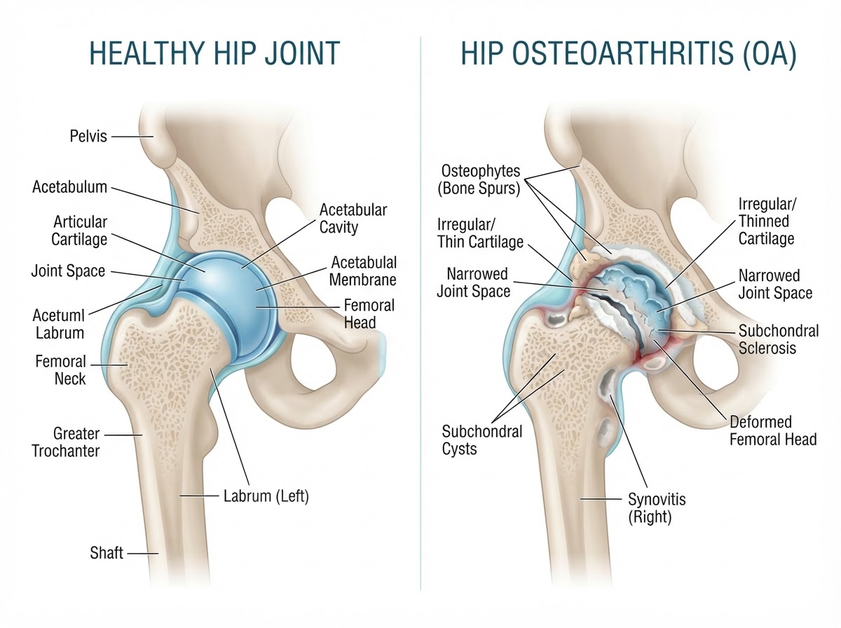
The hip is a ball-and-socket joint: the ball is the femoral head and the socket is the acetabulum of the pelvis. In a healthy hip, both surfaces are covered by articular cartilage, a smooth tissue that allows near-frictionless movement, and the joint space between them is maintained by that cartilage. In osteoarthritis the cartilage thins and is progressively lost, the joint space narrows, and the subchondral bone beneath the cartilage reacts by becoming denser and harder (subchondral sclerosis). The synovium, the thin lining of the joint, may become inflamed and produce excess fluid. As the joint remodels, osteophytes — bony outgrowths at the joint margins — form, and the femoral head may gradually change shape. These structural changes explain the stiffness, pain and reduced range of motion that patients experience, though their severity on imaging does not always match the intensity of symptoms.
Common symptoms of hip osteoarthritis
The most characteristic symptom is groin pain, often radiating towards the front of the thigh and sometimes towards the knee, which can occasionally be the dominant complaint and mislead the patient into thinking the problem is in the knee. Stiffness is typical, especially after rest or first thing in the morning, and a reduced range of motion — particularly internal rotation — is an early sign. Many patients notice difficulty putting on shoes and socks, getting up from a low chair or getting in and out of a car. Pain when walking, especially as distance increases, and difficulty starting to walk after a period of sitting are common. In more advanced cases, night pain can disturb sleep, particularly when lying on the affected side. Symptoms vary considerably between individuals, and two people with similar X-rays can have very different levels of pain and function.
Why does hip osteoarthritis develop?
Hip osteoarthritis is usually the result of several factors acting together. Age-related degeneration of cartilage is the most common contributor, and genetic predisposition influences how an individual's joints age. Previous trauma to the hip, such as a fracture or a labral injury, can accelerate wear. Developmental hip disorders and femoroacetabular impingement (FAI) — where an altered shape of the femoral head or acetabulum creates abnormal contact during movement — can predispose the joint to early osteoarthritis by concentrating load over a smaller area. Previous hip surgery, obesity and sustained mechanical loading also contribute, and secondary osteoarthritis can follow specific conditions such as avascular necrosis or inflammatory arthritis. Understanding the underlying cause matters because it can influence treatment: a young patient with impingement and early cartilage change has a different pathway from an older patient with advanced degenerative disease.
How is hip osteoarthritis diagnosed?
Diagnosis combines medical history, clinical examination and imaging, interpreted together rather than in isolation. The history establishes the pattern of pain, its effect on daily life and the patient's functional demands. Examination assesses range of motion — loss of internal rotation is an early hip sign — gait, leg length and pain on specific movements. Plain radiographs remain the first-line imaging test: a weight-bearing anteroposterior pelvis view and a lateral hip view show joint-space narrowing, osteophytes, sclerosis and femoral head shape. MRI is not routinely required for straightforward osteoarthritis but is useful when the diagnosis is uncertain, when younger patients have atypical pain, or when labral, cartilage or bone pathology such as avascular necrosis is suspected. A differential diagnosis is important because pain referred from the lumbar spine, trochanteric pain, hip flexor tendinopathy and knee pathology can all mimic hip arthritis. Imaging findings must always be interpreted alongside symptoms and examination.
Understanding hip osteoarthritis severity
Hip osteoarthritis is broadly described as mild, moderate or advanced. Radiographic features include joint-space narrowing (the most reliable indicator of cartilage loss), osteophytes at the joint margins, subchondral sclerosis, subchondral cysts and progressive deformity of the femoral head. A key point is that radiographic severity does not always correspond directly to pain intensity: some patients with severe X-ray changes have modest symptoms and remain active, while others with milder imaging have significant pain and limitation. This is why severity is a clinical judgement that integrates the imaging with the patient's symptoms, function and goals, rather than a grade read off an X-ray. Treatment decisions follow that integrated assessment, not the radiograph alone.
Conservative treatment options
Conservative management is the foundation of care for most patients, particularly in early and moderate disease. Exercise therapy — including strengthening of the hip and core muscles, mobility work and cardiovascular conditioning — is strongly supported by guidelines and helps maintain function and reduce pain. Physiotherapy guides a progressive, individualised programme. Weight management reduces the mechanical load on the hip and is one of the most effective conservative measures. Activity modification — pacing activities, avoiding prolonged standing and using sensible ergonomics — helps patients stay active within their limits. Walking aids, when appropriate, reduce load and improve confidence. Pain management, including simple analgesia and anti-inflammatics used judiciously, supports engagement with exercise. The OARSI and NICE guidelines emphasise that conservative care should be optimised and sustained, and that it remains relevant even when surgery is being considered.
Injections and biological treatments
Injections can provide symptom relief in selected patients but have important limitations. Corticosteroid injections can offer short-term reduction in pain and inflammation, particularly when synovitis is prominent, but their benefit is often transient and repeated injections are not advised. Hyaluronic acid injections have been used for hip osteoarthritis, though the evidence is less consistent than for the knee and guidelines vary in their recommendation. Platelet-rich plasma (PRP) and other orthobiological treatments are areas of active research, but the evidence for hip osteoarthritis is still emerging and less established than for some knee applications; they should not be presented as proven cartilage regeneration. It is important to distinguish established treatments from interventions with uncertain or emerging evidence, and to be clear that no injection has been shown to regenerate hip cartilage or reverse established osteoarthritis. Injections are best seen as one option within an individualised plan, used when clinically appropriate and never as a substitute for exercise, weight management and activity modification.
Hip preservation: can joint replacement be delayed?
Hip preservation refers to joint-preserving procedures aimed at addressing an underlying structural abnormality before advanced arthritis develops. It is most relevant for younger patients with early-stage disease and a correctable problem such as femoroacetabular impingement, where reshaping the bone can reduce abnormal contact and may slow progression. Patient selection is critical: preservation is considered for appropriate patients with early disease and a defined structural abnormality, not for advanced osteoarthritis where the cartilage is already lost. In established arthritis, joint-preserving surgery has limited value and may even worsen symptoms. This is why not every patient is a candidate for hip preservation, and why the decision depends on a careful assessment of the anatomy, the stage of the disease and the patient's goals. For many patients with moderate symptoms, optimised conservative care — rather than surgery — is the most effective way to delay a replacement.
When is hip replacement recommended?
Hip replacement is considered when persistent pain and functional limitation persist despite appropriate conservative treatment and begin to affect daily life. Typical indications include pain that limits walking, disturbs sleep or restricts independence; stiffness that interferes with dressing, driving or rising from a chair; and failure of an adequate trial of conservative care. The decision also weighs the clinical and radiographic assessment, the patient's age, activity and expectations, and the expected durability of the implant. Crucially, surgery is not recommended solely on the basis of an X-ray: a patient with severe radiographic changes but manageable symptoms may not need surgery, while a patient with moderate imaging but marked functional limitation may. The decision is a shared one, made after understanding what the patient wants to achieve and what the operation can realistically deliver.
Modern hip replacement surgery
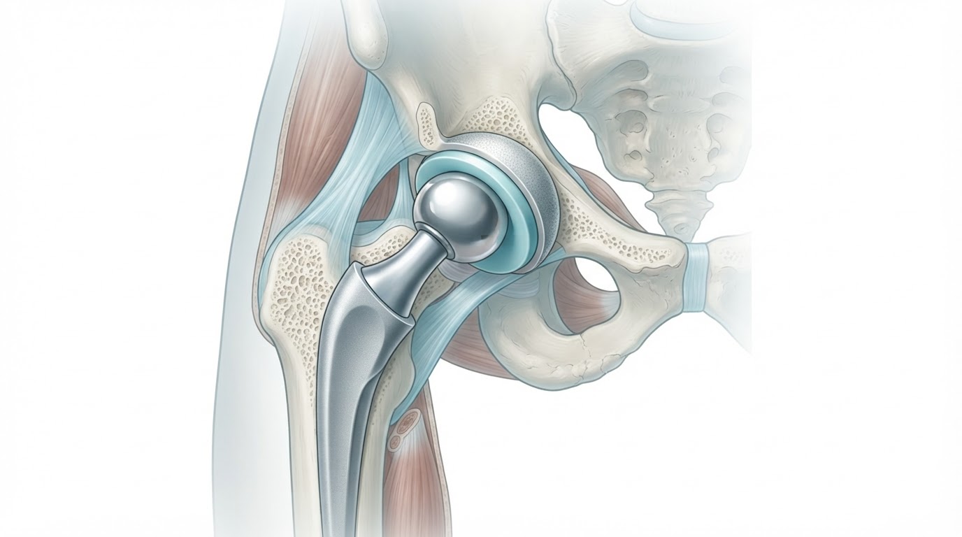
Total hip arthroplasty replaces the damaged articulation with an acetabular component placed in the pelvis and a femoral stem inserted into the femur, topped by a prosthetic femoral head that articulates against a bearing surface in the acetabular component. Implant fixation can be cemented or uncemented depending on bone quality and the patient, and the bearing surface — metal, ceramic or highly cross-linked polyethylene — is selected individually. Several surgical approaches exist, including posterior, anterolateral and direct anterior approaches, each with its own characteristics in terms of muscle-sparing, recovery and precautions. Minimally invasive approaches can reduce early discomfort and speed early mobilisation, but no single approach is universally superior; the choice depends on the surgeon's experience, the patient's anatomy and the individual situation. Individualised surgical planning — including implant selection, leg-length management and approach — is central to a good outcome and a predictable functional recovery.
Recovery after hip replacement
Recovery after hip replacement is generally faster than many patients expect. Early mobilisation — often on the day of surgery or the day after — is encouraged, with walking progression using aids as needed. Rehabilitation focuses on restoring mobility, strength and confidence, and most patients return to daily activities within weeks. Driving can usually resume once strength and reflexes allow safe control, typically several weeks depending on the side and the approach. Return to work depends on the demands of the job: desk-based work can resume relatively quickly, while physically demanding work takes longer. Long-term expectations are generally very good: most patients experience substantial pain relief and a marked improvement in function and quality of life, and modern implants can last many years, though no specific recovery time or implant lifespan can be guaranteed for an individual. Recovery varies according to the approach, the patient's fitness and any complications, so milestones are set after assessment rather than promised in advance.
Choosing the right treatment
The table below is an educational framework, not an automatic treatment algorithm. It illustrates how the approach may differ according to the clinical situation, but every decision is individualised after assessment. | Clinical situation | Possible treatment approach | |---|---| | Mild disease | Conservative treatment | | Persistent symptoms | Individualised assessment and additional treatment | | Selected structural abnormalities | Consideration of hip preservation | | Advanced symptomatic disease | Assessment for hip replacement | The aim is to match the treatment to the patient's symptoms, anatomy, functional needs and expectations, and to revisit that decision over time as symptoms and circumstances change.
Conclusion
Hip osteoarthritis is a progressive but manageable condition. Early assessment, accurate diagnosis and treatment selected according to the patient's symptoms, anatomy, functional needs and expectations are the keys to a good outcome. Conservative care — exercise, weight management and activity modification — remains the foundation for most patients; injections can help in selected cases but do not regenerate cartilage; hip preservation is appropriate for carefully selected patients with early disease and a correctable abnormality; and modern hip replacement offers excellent pain relief and function for advanced symptomatic disease. No decision should be made on the basis of an X-ray alone. A consultation with an orthopaedic surgeon is the way to determine which approach is right for your hip.
The Bottom Line: Matching Treatment to the Hip
Hip osteoarthritis is managed, not simply operated on. Conservative care is the foundation for most patients, injections help in selected cases without regenerating cartilage, hip preservation suits carefully selected early disease, and modern hip replacement offers excellent relief for advanced symptomatic arthritis. Treatment is individualised after a specialist assessment that integrates symptoms, examination and imaging — never an X-ray alone. A consultation with an orthopaedic surgeon is the way to decide what is appropriate for your hip.
References & scientific evidence
- Osteoarthritis in over 16s: assessment and management. NICE guideline NG226.. National Institute for Health and Care Excellence (NICE), 2022.Clinical guidelineView source
- Management of Osteoarthritis of the Hip: Evidence-Based Clinical Practice Guideline.. American Academy of Orthopaedic Surgeons (AAOS), 2017.Clinical guidelineView source
- OARSI guidelines for the non-surgical management of knee, hip, and polyarticular osteoarthritis.. Osteoarthritis and Cartilage, 2013.Clinical guidelineDOI: 10.1016/j.joca.2013.02.013View source
- 2019 American College of Rheumatology/Arthritis Foundation Guideline for the Management of Osteoarthritis of the Hand, Hip, and Knee.. Arthritis Care & Research, 2020.Clinical guidelineDOI: 10.1002/acr.24131PMID: 31908149View source
- Bannuru RR et al.. Effectiveness of pharmacologic and non-pharmacologic treatments for knee and hip osteoarthritis: a systematic review and meta-analysis.. Osteoarthritis and Cartilage, 2019.Meta-analysisDOI: 10.1016/j.joca.2019.06.011View source
- Hochberg MC et al.. When is osteoarthritis of the hip or knee an appropriate indication for joint replacement? A systematic review of the literature.. BMJ Open, 2012.Systematic reviewDOI: 10.1136/bmjopen-2012-000631View source
Frequently asked questions

Dr. Pablo Banchero Lasalvia
Orthopaedic Surgeon · Traumatology Team
- Author:
- Dr. Pablo Banchero Lasalvia
- Medical reviewer:
- Dr. Pablo Banchero Lasalvia
- Published:
- 1 November 2026
- Medical review date:
- 1 November 2026
- ·
- 14 min read
Book Your Consultation via WhatsApp
Request a private orthopaedic assessment with Dr Pablo Banchero on the Costa Blanca.
Official number: +34 602 99 22 22