
Hip arthroscopy is a minimally invasive procedure that allows a surgeon to see inside the hip joint and treat certain problems through small incisions. It can be an excellent option for the right patient — typically someone with a treatable intra-articular problem such as femoroacetabular impingement or a labral tear, preserved joint space and limited degeneration. But it is not a treatment for established advanced hip osteoarthritis, and for some patients it is simply too late for arthroscopy to help. The central clinical question this article answers is: who is a good candidate for hip arthroscopy, and when is it too late to preserve the joint with this procedure? It explains how the operation works, what femoroacetabular impingement and labral tears are, how patient selection is made, what recovery involves, the complications that can occur, what the scientific evidence shows about outcomes and conversion to hip replacement, and how the decision is individualised. It is written for residents and visitors on the Costa Blanca — including patients in Alicante, Torrevieja and Orihuela Costa — and for international patients seeking a second opinion about joint preservation. The aim is to help you understand honestly whether arthroscopy is appropriate for your hip, not to promote it as a universal solution.
Introduction: what is hip arthroscopy?
Hip arthroscopy is a minimally invasive surgical technique in which a small camera (the arthroscope) and fine instruments are inserted into the hip joint through a few small portals, allowing the surgeon to visualise and treat certain intra-articular problems without a large open incision. It is used most often for femoroacetabular impingement, labral tears and selected chondral (cartilage) injuries. It is important to be clear from the outset about what hip arthroscopy is not: it is not a treatment for established advanced hip osteoarthritis, it cannot regrow lost cartilage, and it is not appropriate for every painful hip. The value of arthroscopy depends entirely on selecting the right patient with the right pathology at the right stage of disease. This article focuses on that selection — who benefits, who does not, and where the line falls.
How hip arthroscopy is performed
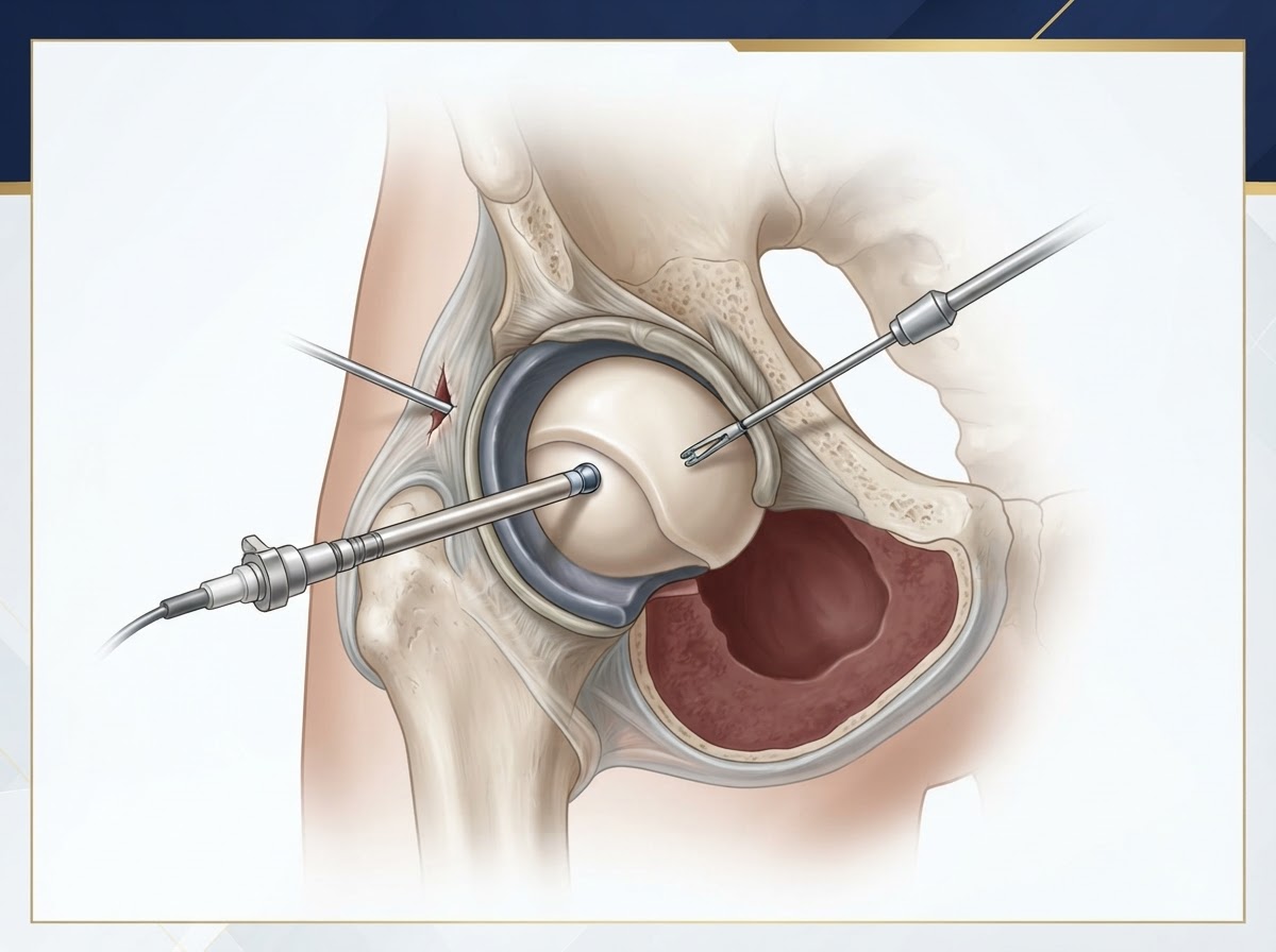
Hip arthroscopy is performed under general or regional anaesthesia. Because the hip joint is deep and tightly enclosed, the joint must first be distracted — gently pulled apart using traction on a specialised operating table — to create enough space to insert the instruments safely. Small portals (typically two or three) are made at the side of the hip, the arthroscope is introduced, and the joint is filled with fluid to distend it and improve visibility. The surgeon inspects the central and peripheral compartments of the joint, identifies the pathology, and performs the planned procedures — which may include reshaping bone (femoroplasty for a cam lesion, acetabular rim trimming for a pincer lesion), repairing or debriding the labrum, treating focal cartilage damage, and managing the capsule. The procedure usually takes between one and two hours. Traction is released as soon as it is safe to do so, and the portals are closed with small sutures.
Hip anatomy: labrum, cartilage, capsule and femoral head
Understanding the procedure requires a basic grasp of hip anatomy. The hip is a ball-and-socket joint: the ball is the femoral head, and the socket is the acetabulum, which is deepened by a fibrocartilaginous ring called the labrum. The labrum acts as a seal and stabiliser, distributing load and joint fluid and helping to maintain negative pressure that stabilises the joint. Both the femoral head and the acetabulum are covered by articular cartilage, the smooth tissue that allows low-friction movement. The joint is enclosed by a fibrous capsule reinforced by ligaments, which provides stability. Hip arthroscopy works by addressing problems within these structures — most commonly a damaged labrum, abnormal bone morphology that causes impingement, and focal cartilage injury — while preserving the normal anatomy where possible.
Femoroacetabular impingement: CAM morphology
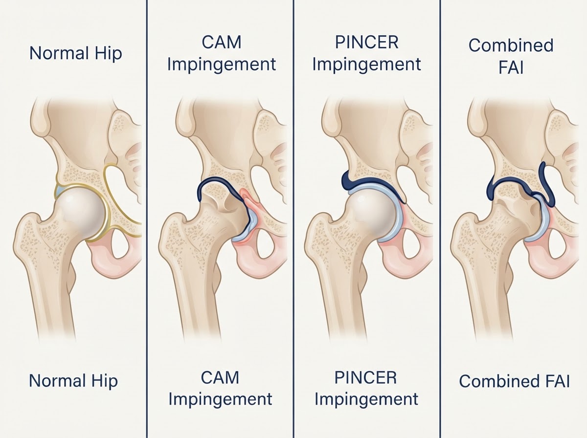
Femoroacetabular impingement (FAI) occurs when the shape of the femoral head–neck junction or the acetabulum causes abnormal contact during hip movement, damaging the labrum and cartilage over time. In CAM morphology, there is an excess of bone (a 'bump') on the femoral head–neck junction, so the head is not perfectly round. As the hip flexes and rotates, this bony prominence is driven into the acetabular rim, shearing the labrum and cartilage. CAM morphology is more common in young and middle-aged men and is frequently associated with labral tears and chondral damage at the anterosuperior acetabulum. It is important to understand that a cam morphology seen on imaging is common and does not automatically mean surgery is necessary: many people have cam morphology and no symptoms. Surgery is considered when symptoms, clinical examination and imaging are consistent and appropriate conservative treatment has not provided sufficient improvement.
Femoroacetabular impingement: PINCER morphology
In PINCER morphology, the acetabulum covers (or 'over-covers') the femoral head more than usual, so the rim of the socket presses against the femoral neck during movement. This can occur because the acetabulum is too deep (coxa profunda or protrusio) or because it is retroverted (turned backwards). The repetitive contact primarily damages the labrum, which may become crushed or torn, and in some cases can lead to cartilage injury. Pincer morphology is more common in middle-aged women. As with cam morphology, the presence of pincer morphology on imaging alone is not an indication for surgery; it must be correlated with symptoms and examination. When surgery is appropriate, the treatment is careful acetabular rim trimming to reduce the over-coverage — but excessive resection can create instability, so preservation of normal anatomy and careful judgement are essential.
Combined CAM and PINCER morphology
In many patients, both cam and pincer morphology coexist — this is called combined or mixed impingement and is in fact the most common pattern. The combined mechanism tends to cause more extensive labral and chondral damage than either alone, because both the femoral bump and the over-covering rim contribute to abnormal contact. Surgical treatment addresses both components: femoroplasty to reshape the femoral head–neck junction and acetabular rim trimming to reduce over-coverage, combined with labral repair or debridement as indicated. The presence of combined morphology does not by itself determine the need for surgery; the decision rests, as always, on the correlation of symptoms, examination and imaging, and on the failure of appropriate non-operative management.
Labral tears: symptoms, diagnosis and treatment
The acetabular labrum can tear traumatically (for example, in sport or a dislocation) or degeneratively (in association with impingement or osteoarthritis). A labral tear may cause groin pain, mechanical symptoms such as clicking, locking or catching, a sensation of giving way, and pain on flexion and rotation. Diagnosis combines clinical examination (provocative tests such as the FADIR test) with imaging: MRI is useful, and MR arthrography (MRI with contrast injected into the joint) has higher sensitivity for labral tears. Not every labral tear detected on MRI requires surgery — many tears are incidental or degenerative and may be managed conservatively, especially when there is no significant mechanical symptom or when degenerative change is present. When surgery is indicated, labral repair (reattaching the labrum) is preferred over debridement (trimming it away) whenever the tissue is repairable, because preserving the labrum is important for joint function. Associated cartilage damage and the underlying impingement must be addressed at the same time, as they strongly influence outcomes.
Chondral injuries and cartilage damage
Chondral injuries are injuries to the articular cartilage that covers the femoral head and acetabulum. They range from softening and superficial fissuring to full-thickness cartilage loss exposing the underlying bone. A focal chondral injury — a discrete area of damage in an otherwise healthy joint — is very different from diffuse degenerative cartilage loss, which is a feature of osteoarthritis. This distinction is critical for arthroscopy: a focal lesion in a patient with preserved joint space may be treatable, while diffuse cartilage loss indicates established osteoarthritis in which arthroscopy is generally not helpful. The severity and location of chondral damage are among the strongest predictors of outcome after hip arthroscopy, and significant chondral damage — particularly full-thickness loss — is associated with poorer results and a higher likelihood of later hip replacement.
Other indications for hip arthroscopy
Beyond femoroacetabular impingement and labral tears, hip arthroscopy may be used for selected other indications, including the removal of loose bodies (fragments of bone or cartilage floating in the joint that cause locking), synovial disorders (such as synovial chondromatosis), biopsy of unclear joint pathology, the drainage and washout of a septic hip in selected cases, and certain extra-articular problems such as iliopsoas tendon release or snapping hip syndromes in carefully selected patients. These indications are more limited and specific than the management of impingement, and each carries its own selection criteria. The common principle is that arthroscopy is appropriate when there is a defined, treatable problem that is likely to benefit from a minimally invasive approach, and when the joint is not already arthritic.
Clinical assessment and diagnostic imaging
A thorough assessment is the foundation of good patient selection. The clinical history establishes the pain pattern, mechanical symptoms, activity limitation and any history of hip problems in childhood (such as dysplasia, Perthes or slipped capital femoral epiphysis). Physical examination assesses range of motion and performs provocative tests for impingement and labral pathology. Imaging is then used to confirm the diagnosis and assess the joint. Weight-bearing anteroposterior pelvic and hip radiographs are essential to evaluate joint-space width, osteoarthritis grade, femoral head shape, acetabular coverage and any dysplasia. Cross-sectional imaging — MRI, MR arthrography and sometimes CT — is used to assess the labrum, cartilage, and bone morphology in detail. Crucially, imaging findings must be correlated with symptoms and examination: imaging alone never dictates surgery, and asymptomatic cam or pincer morphology is common.
MRI, MR arthrography and CT: when are they useful?
MRI is useful for assessing the labrum, cartilage, synovium and surrounding soft tissues, and for excluding other causes of hip pain. MR arthrography — MRI after injecting contrast into the joint — has higher sensitivity for labral tears and is often preferred when labral pathology is the main question. CT provides detailed assessment of bone morphology and is particularly useful for measuring cam lesions, acetabular version and coverage, and for planning bone reshaping; three-dimensional CT reconstructions can help the surgeon plan the femoroplasty. CT is also valuable in the assessment of component rotation and bone defects in the context of a painful prosthesis, though that is a separate scenario. None of these investigations is routine for every patient; they are selected according to the clinical question. The goal is to build a coherent picture in which symptoms, examination and imaging agree before surgery is recommended.
Tönnis classification and hip osteoarthritis
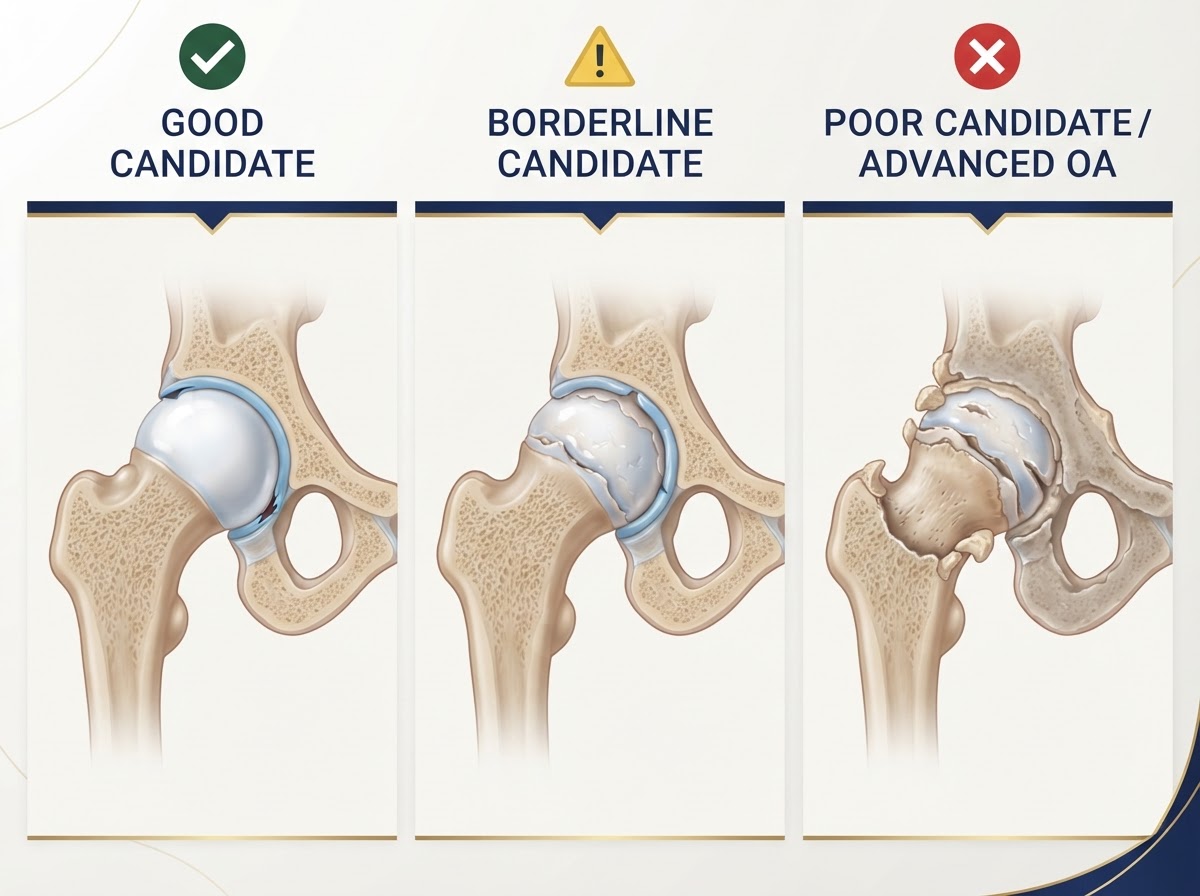
The Tönnis classification grades hip osteoarthritis on radiographs from grade 0 (no osteoarthritis) to grade 3 (severe osteoarthritis). Grade 0 shows no arthritic change; grade 1 shows increased sclerosis and slight joint-space narrowing with minor osteophytes; grade 2 shows small cysts, moderate joint-space narrowing and moderate osteophytes; grade 3 shows large cysts, severe joint-space narrowing, marked osteophytes and structural changes. The Tönnis grade is a key factor in arthroscopy candidacy: lower grades (0 and 1) are generally associated with better outcomes, while higher grades (2 and especially 3) are associated with poorer outcomes and a higher rate of conversion to hip replacement. The Kellgren-Lawrence classification is another widely used system that grades osteoarthritis from 0 to 4. These grading systems help frame the decision, but they are interpreted alongside joint-space width, chondral damage, symptoms and the overall clinical picture — a grade alone never determines treatment.
Joint-space width: why the 2 mm threshold matters
The minimum joint-space width, measured on a weight-bearing radiograph, is one of the most useful single predictors of outcome after hip arthroscopy. A joint space of approximately 2 mm or less has been associated in several observational studies with a higher likelihood of poor outcomes and a higher rate of conversion to total hip replacement after arthroscopy. The reason is intuitive: a narrow joint space reflects significant cartilage loss, and arthroscopy cannot replace lost cartilage. However, the 2 mm figure must be understood correctly. It is not an absolute universal contraindication — it is a recognised threshold that must be interpreted alongside age, the Tönnis grade, the extent of chondral damage, the presence of subchondral changes, and the overall clinical picture. A borderline patient with a clear, treatable mechanical problem and realistic expectations may still be considered by an experienced hip preservation surgeon, while a patient well below 2 mm with established osteoarthritis is generally a poor candidate. The threshold is a strong guide, not a rigid rule.
Patient selection: who is a good candidate?
A good candidate for hip arthroscopy typically has several features in combination: persistent symptoms compatible with intra-articular pathology (groin pain, mechanical symptoms); positive clinical examination findings (impingement or labral tests); confirmed treatable pathology on imaging (impingement morphology, a repairable labral tear, a focal chondral lesion); preserved joint space; limited degenerative cartilage damage; realistic expectations about what surgery can achieve; and failure of appropriate non-operative treatment (activity modification, physiotherapy, analgesia, and sometimes injections). The interaction of these factors matters more than any single one. A patient with classic impingement symptoms, a repairable labral tear, preserved joint space and no osteoarthritis is an excellent candidate; a patient with the same symptoms but advanced joint-space narrowing and diffuse cartilage loss is not. Selection is individualised and is the single most important determinant of outcome.
When hip arthroscopy has a poor prognosis
Hip arthroscopy has a poor prognosis when the joint is already significantly arthritic. Specific features that predict poor outcomes include advanced Tönnis grade (especially grade 2 or 3), severe joint-space narrowing (commonly around 2 mm or less, interpreted alongside other factors), diffuse cartilage loss or full-thickness chondral defects, subchondral cysts and bone-marrow oedema, significant osteophytes causing mechanical restriction, severe stiffness, advanced femoral head or acetabular degeneration, and older age in combination with degenerative findings. In these situations arthroscopy is unlikely to provide lasting benefit and may, in some cases, worsen symptoms or accelerate the need for hip replacement. The honest message is that performing arthroscopy in advanced osteoarthritis can delay definitive treatment without providing meaningful long-term benefit — and that delay is not always harmless.
Age, body weight and other prognostic factors
Age alone should not determine suitability for hip arthroscopy — a younger patient with advanced degenerative change may be a poor candidate, while an older patient with preserved joint space and a clear, treatable mechanical problem may do well. What matters is the combination of age with the radiographic and chondral findings. Higher body weight and obesity are associated with somewhat poorer outcomes and a higher rate of conversion to hip replacement in some studies, and weight management is part of optimising any candidate. Other factors that influence prognosis include the severity of chondral damage, the presence of subchondral changes, whether the labrum is repairable, the degree of preoperative stiffness, and the patient's activity expectations. No single factor is decisive; the surgeon weighs them together to estimate the likelihood of a meaningful, durable benefit.
Surgical techniques and treatment options
The principal techniques used during hip arthroscopy include: femoral osteochondroplasty (reshaping the femoral head–neck junction to remove a cam lesion and restore a smooth, spherical head); acetabular rim trimming in carefully selected pincer cases to reduce over-coverage (with care to avoid excessive resection that could create instability); labral repair, reattaching the labrum to preserve its function whenever the tissue is repairable; labral debridement, trimming irreparable or degenerative tissue when repair is not possible; capsular management and repair, closing the capsule at the end of the procedure to restore stability; and treatment of focal chondral lesions where indicated. These techniques are tailored to the individual pathology. It is important to state that cartilage restoration procedures are not established solutions for diffuse advanced hip osteoarthritis, and that excessive acetabular rim resection can create instability — preservation of normal anatomy is a core principle.
Rehabilitation and recovery
Recovery after hip arthroscopy depends on the procedures performed and the patient's condition, so fixed recovery guarantees are not appropriate. Early postoperative protection may include restrictions on weight-bearing (often partial weight-bearing with crutches for a few weeks, depending on the extent of bone reshaping and labral repair), limitations on certain movements (avoiding extremes of flexion, rotation or extension for a period), and sometimes a brace. Physiotherapy is central and progresses in stages: early range-of-motion exercises to prevent stiffness, then gradual strengthening of the hip and core muscles, restoration of normal movement patterns, and a controlled return to activity. Return to driving, work and sport depends on the procedure, the side operated, and progress in rehabilitation — driving may resume once off strong analgesia and able to control the vehicle safely, desk work within a couple of weeks, and return to sport over several months. Factors influencing recovery include the extent of surgery, the patient's preoperative condition, adherence to rehabilitation, and individual healing.
Complications and possible adverse outcomes
Hip arthroscopy is generally safe in experienced hands, but complications can occur. Traction-related neuropraxia (temporary nerve irritation from the traction used to distract the joint) can affect the pudendal nerve or other nerves, causing numbness or sensory changes that usually resolve but can persist. Pudendal nerve symptoms and perineal soft-tissue complications are related to traction pressure and duration. Heterotopic ossification (abnormal bone formation in soft tissues) can occur and is reduced by careful technique and sometimes prophylaxis. Infection is uncommon but possible. Fracture of the femoral neck is a rare but serious risk, particularly after extensive cam resection. Persistent pain can occur, especially when patient selection was suboptimal. Capsular instability can result from inadequate capsular closure. Iatrogenic cartilage injury can occur from instrument passage. Some patients ultimately require further surgery, including conversion to hip replacement. Appropriate traction management and careful surgical technique are important in minimising these risks.
Conversion to total hip replacement after arthroscopy
A proportion of patients who undergo hip arthroscopy later require total hip replacement. The reported rates and time to conversion vary considerably between studies and depend on the population, the severity of pre-existing osteoarthritis, the joint-space width, the Tönnis grade, the extent of chondral damage, the patient's age and the length of follow-up. Studies consistently show that conversion risk is higher in patients with more advanced osteoarthritis, lower joint-space width (notably around 2 mm or less), higher Tönnis grade, older age and more extensive chondral damage, and lower in patients with preserved joint space and minimal degeneration. It is important to interpret conversion figures according to the population and follow-up period in each study — a short follow-up will understate the lifetime conversion risk, and a population selected for early disease will show lower rates than one including degenerative cases. The good news is that total hip replacement after previous arthroscopy generally remains effective, though prior surgery can make the subsequent operation somewhat more complex in some cases.
Hip arthroscopy versus total hip replacement
Hip arthroscopy and total hip replacement address different problems and are not interchangeable. Arthroscopy aims to treat a specific intra-articular problem — most often impingement with labral pathology — in a joint that is otherwise healthy, with the goal of relieving symptoms and potentially slowing progression. Total hip replacement replaces the arthritic joint surfaces and is the definitive treatment for advanced, symptomatic osteoarthritis. The two procedures sit at opposite ends of the disease spectrum: arthroscopy is for the preserved joint with a focal problem; replacement is for the arthritic joint. The error to avoid is offering arthroscopy to a patient whose joint is already arthritic in the hope of avoiding a replacement — in that situation arthroscopy is unlikely to help and may delay appropriate definitive treatment. The decision between the two is based on the stage of disease, the pathology present, the patient's symptoms and expectations, and shared decision-making with an experienced surgeon.
Conclusion and individualised treatment planning
Hip arthroscopy is a valuable procedure for the right patient: someone with a treatable intra-articular problem such as femoroacetabular impingement or a labral tear, preserved joint space and limited degeneration, in whom appropriate conservative treatment has not provided sufficient relief. It is not a treatment for established advanced hip osteoarthritis, and for patients with significant joint-space narrowing, high Tönnis grade, diffuse cartilage loss or extensive degenerative change, it is generally too late for arthroscopy to provide lasting benefit. The 2 mm joint-space threshold is a strong guide but not an absolute rule, and age alone does not determine suitability — the decision is individualised, weighing symptoms, examination, imaging, chondral status and expectations together. For residents and visitors across the Costa Blanca — Alicante, Torrevieja and Orihuela Costa — Dr Pablo Banchero offers individualised, evidence-based assessment and an honest second opinion to help you decide whether hip arthroscopy is appropriate for you, or whether conservative management or hip replacement is the better path.
The right procedure for the right hip, at the right time
Hip arthroscopy can provide excellent outcomes in well-selected patients with femoroacetabular impingement, labral tears and focal pathology in a preserved joint, but it is not a treatment for advanced hip osteoarthritis. Joint-space width of around 2 mm or less, high Tönnis grade, diffuse cartilage loss and extensive degenerative change predict poorer outcomes and a higher rate of conversion to hip replacement — and arthroscopy in advanced disease can delay appropriate definitive treatment without meaningful benefit. The decision is individualised, based on the correlation of symptoms, examination and imaging, and on shared decision-making. On the Costa Blanca — Alicante, Torrevieja and Orihuela Costa — Dr Pablo Banchero provides evidence-based assessment and second opinions on whether hip arthroscopy, conservative management or hip replacement is the right choice for your hip.
References & scientific evidence
- Griffin DR, Dickenson EJ, O'Donnell J, et al.. The Warwick Agreement on femoroacetabular impingement syndrome (FAI syndrome): an international consensus statement.. British Journal of Sports Medicine, 2016.Clinical guidelineDOI: 10.1136/bjsports-2016-096743View source
- Domb BG, Martin TJ, Gu C, et al.. Predictors of clinical outcomes after hip arthroscopy: a 5-year prospective multicenter study.. American Journal of Sports Medicine, 2020.Observational studyDOI: 10.1177/0363546520909333View source
- Maldonado DR, Chen AW, Lall AC, et al.. Hip arthroscopy in patients with Tönnis grade 2 osteoarthritis: outcomes and conversion to total hip arthroplasty.. Journal of Arthroplasty, 2021.Observational studyDOI: 10.1016/j.arth.2021.02.038View source
- Degen RM, Pan TJ, Chang B, et al.. Risk of total hip arthroplasty after hip arthroscopy in patients with pre-existing osteoarthritis.. Journal of Hip Preservation Surgery, 2018.Observational studyDOI: 10.1093/jhps/hny015View source
- Kunze KN, Beck EC, Nwachukwu BU, et al.. Early hip arthroscopy for femoroacetabular impingement: a systematic review of outcomes and complications.. Arthroscopy, 2019.Systematic reviewDOI: 10.1016/j.arthro.2019.07.020View source
- Mansell JP, Khanna V, Bhatia S, et al.. Femoroacetabular impingement: a comparison of hip arthroscopy and non-operative management — a randomised controlled trial.. British Journal of Sports Medicine, 2023.Randomised controlled trialDOI: 10.1136/bjsports-2022-106029View source
- Espinosa C, Khanduja V.. Arthroscopic management of femoroacetabular impingement: a systematic review of mid-term outcomes.. Journal of Hip Preservation Surgery, 2020.Systematic reviewDOI: 10.1093/jhps/hnaa018View source
- Haviv B, O'Donnell J, Takla A, et al.. Arthroscopic femoral osteochondroplasty for cam-type femoroacetabular impingement: outcomes and predictors of conversion to total hip arthroplasty.. Journal of Arthroplasty, 2018.Observational studyDOI: 10.1016/j.arth.2018.01.052View source
- Chandrasekaran S, Darwish N, Close M, et al.. Minimum 5-year outcomes following hip arthroscopy in patients with Tönnis grade 2 versus grade 0 or 1 osteoarthritis.. Journal of Arthroscopy and Related Surgery, 2019.Observational studyDOI: 10.1016/j.arthro.2019.04.020View source
- Philippon MJ, Briggs KK, Carlisle JC, Patterson DC.. Joint space predicts THA after hip arthroscopy in patients 50 years and older.. Clinical Orthopaedics and Related Research, 2013.Observational studyDOI: 10.1007/s11999-012-2740-2View source
- Domb BG, Gui C, Lodhia P.. How much arthritis is too much for hip arthroscopy: a systematic review of outcomes in patients with Tönnis grade 2 osteoarthritis.. Arthroscopy, 2015.Systematic reviewDOI: 10.1016/j.arthro.2015.02.042View source
- Weber AE, Kuhns BD, Bhatia S, et al.. Hip arthroscopy in the older patient: outcomes and predictors of conversion to total hip arthroplasty.. Arthroscopy, 2017.Observational studyDOI: 10.1016/j.arthro.2016.10.014View source
- Tönnis D, Heinecke A.. Acetabular and femoral anteversion: relationship with osteoarthritis of the hip.. Journal of Bone and Joint Surgery (American), 1999.Observational studyDOI: 10.2106/00004623-199907000-00004View source
- American Academy of Orthopaedic Surgeons (AAOS). Management of Osteoarthritis of the Hip — Evidence-Based Clinical Practice Guideline.. AAOS Clinical Practice Guidelines, 2023.Clinical guidelineView source
- National Joint Registry (NJR) for England, Wales, Northern Ireland and the Isle of Man. National Joint Registry 21st Annual Report — Hip, Knee, Ankle, Elbow and Shoulder Arthroplasty: Outcomes and Survivorship.. National Joint Registry, 2024.Observational studyView source
Frequently asked questions

Dr. Pablo Banchero Lasalvia
Orthopaedic Surgeon · Traumatology Team
- Author:
- Dr. Pablo Banchero Lasalvia
- Medical reviewer:
- Dr. Pablo Banchero Lasalvia
- Published:
- 2 November 2026
- Medical review date:
- 2 November 2026
- ·
- 24 min read
Book Your Consultation via WhatsApp
Request a private, evidence-based orthopaedic assessment with Dr Pablo Banchero to discuss femoroacetabular impingement, a labral tear, whether hip arthroscopy is appropriate for you, or a second opinion on joint preservation on the Costa Blanca.
Official number: +34 602 99 22 22