
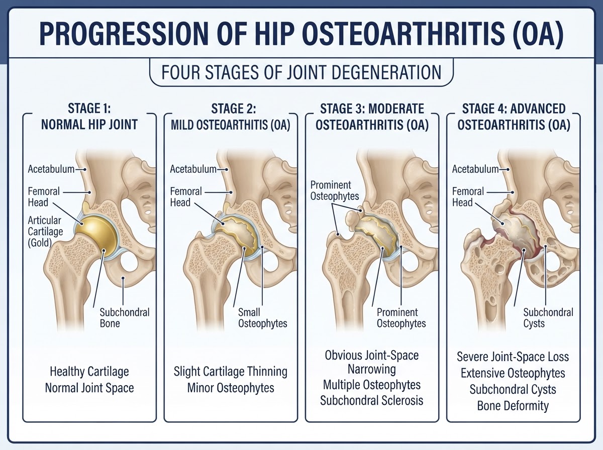
A diagnosis of hip osteoarthritis does not automatically mean a hip replacement. Many patients can be managed for years with exercise, weight management, physiotherapy and carefully chosen injections; some are candidates for joint-preserving procedures; and for a smaller group, total hip replacement becomes the most appropriate and effective option once conservative treatment has been exhausted. The difficulty for patients — and the reason this article exists — is knowing which path applies to them, and when. This article explains, in plain language, what hip osteoarthritis is, how it is graded, what conservative and interventional treatments actually offer (including PRP, hyaluronic acid, radiofrequency and hip arthroscopy), what the current scientific evidence supports and what it does not, and how an orthopaedic surgeon decides, together with the patient, when the time has come to operate. It is written for residents and visitors on the Costa Blanca — including patients in Alicante, Torrevieja and Orihuela Costa — and for international patients seeking a second opinion before committing to surgery. The aim is to help you understand your options honestly, not to push a particular treatment.
Introduction: does every patient with hip osteoarthritis need a replacement?
No. Hip osteoarthritis is common, progressive and variable: some people live with mild changes for decades with little trouble, others deteriorate more quickly, and many never reach the point where surgery is justified. The decision to replace a hip is based on symptoms, function and the failure of appropriate non-operative treatment — not on an X-ray grade alone. A radiograph can look severe while a patient functions well, and conversely a patient can suffer significant pain with only moderate radiographic change. This article walks through the full spectrum of management, from conservative care through interventional options and joint preservation to hip replacement, so that you can understand what each option offers, what the evidence supports, and how the timing of surgery is judged.
What is hip osteoarthritis?
Hip osteoarthritis is a progressive degenerative joint disease in which the articular cartilage that covers the femoral head and the acetabulum (the socket) gradually thins and is lost, accompanied by changes in the underlying bone, the joint capsule and the surrounding soft tissues. It is not simply 'wear and tear' — it is an active process involving cartilage breakdown, bone remodelling, low-grade inflammation and altered joint mechanics. The result is pain, stiffness, reduced range of motion and progressive loss of function. Osteoarthritis is classified as primary (idiopathic, often age-related) or secondary (resulting from a known cause such as developmental dysplasia, femoroacetabular impingement, previous trauma, Perthes disease or slipped capital femoral epiphysis). Understanding whether the osteoarthritis is primary or secondary matters, because secondary causes can sometimes be addressed earlier with joint-preserving surgery before the joint is irreversibly damaged.
Causes and risk factors
Hip osteoarthritis is multifactorial. Established risk factors include increasing age, a family history of osteoarthritis, obesity (which increases both mechanical load and systemic inflammatory signalling), previous hip injury or fracture, developmental conditions such as hip dysplasia and femoroacetabular impingement, repetitive heavy occupational or sporting load, and certain metabolic conditions. Some of these — age and genetics — cannot be changed; others, particularly weight and modifiable loading patterns, can be influenced and are the basis of conservative management. It is important to understand that a risk factor is not a cause in the sense of a single trigger; osteoarthritis reflects the combined effect of many factors over a lifetime, and two people with similar risk profiles may follow very different clinical courses.
Symptoms and clinical presentation
Hip osteoarthritis typically causes pain in the groin, the front of the thigh, the lateral hip or sometimes the buttock, and occasionally pain that is felt mainly in the knee (referred pain). The pain is usually worse with weight-bearing and activity and improves with rest, but as the disease progresses pain may become continuous, including night pain and rest pain that disturbs sleep. Stiffness is common, particularly in the morning or after sitting (the so-called 'start-up' pain), and typically lasts less than 30 minutes in early disease. Patients often notice a progressive loss of movement — difficulty putting on socks and shoes, getting in and out of a car, or climbing stairs — and may develop a limp. As function declines, independence and quality of life are affected. The pattern and severity of symptoms, more than the X-ray appearance, guide treatment decisions.
How hip osteoarthritis is diagnosed
Diagnosis is clinical, supported by imaging. A careful history and physical examination establish the pain pattern, functional limitation, mechanical symptoms and any features suggesting an alternative or additional cause (such as lumbar spine referred pain or trochanteric pain syndrome). Examination assesses range of motion, gait, leg length and provocative tests. Weight-bearing anteroposterior pelvic and hip radiographs are the first-line imaging investigation and usually sufficient to confirm osteoarthritis: the classic findings are joint-space narrowing (often superiorly), osteophytes, subchondral sclerosis and subchondral cysts. MRI is not routinely required for diagnosis but may be used when there is diagnostic uncertainty, when a soft-tissue or labral problem is suspected, or when joint-preserving surgery is being considered and the cartilage and labrum need detailed assessment. Blood tests are not diagnostic of osteoarthritis but may be used to exclude inflammatory arthritis when the presentation is atypical.
Radiological classification and joint-space narrowing
Several grading systems describe the severity of hip osteoarthritis on radiographs. The Kellgren-Lawrence system grades from 0 (normal) to 4 (severe), based on osteophytes, joint-space narrowing, sclerosis and deformity. The Tönnis classification (grades 0 to 3) is widely used in hip preservation and grades joint-space narrowing, head deformity and osteophytes. The most clinically useful single measurement is the minimum joint-space width, measured on a weight-bearing radiograph; a joint space of approximately 2 mm or less is a recognised threshold below which the likelihood of significant cartilage loss and poor outcomes from joint-preserving surgery increases. It is important to emphasise that joint space is one important predictor among several — it is not an absolute universal cut-off, and it must be interpreted alongside symptoms, age, the presence of a treatable intra-articular lesion and the overall clinical picture. A radiographic grade alone never determines treatment.
Mild, moderate and advanced osteoarthritis: how treatment changes
Treatment is staged according to disease severity and, more importantly, symptom burden. In mild osteoarthritis, the emphasis is on conservative management: education, therapeutic exercise, weight management, activity modification and simple analgesia, with interventional injections reserved for selected patients. In moderate osteoarthritis, individualised conservative treatment continues, and image-guided interventions such as PRP or hyaluronic acid may be considered after an honest discussion of the evidence; hip arthroscopy is only appropriate when a separate treatable intra-articular pathology exists and joint degeneration is limited. In advanced osteoarthritis with significant pain and disability, the focus shifts to assessing whether total hip replacement is now indicated, and repeated interventions that only delay appropriate surgery without meaningful benefit should be avoided. The transition between stages is gradual and individual — there is no single moment at which conservative care 'stops working', but there is a point at which continuing ineffective treatment becomes a disservice to the patient.
Conservative management
Conservative management is the foundation of treatment at every stage and remains relevant even when surgery is being considered. Therapeutic exercise — strengthening the muscles around the hip and improving flexibility and balance — has the strongest evidence base of any non-operative intervention and should be started early and maintained. Physiotherapy provides a structured, supervised programme and helps optimise movement patterns. Weight management reduces mechanical load and systemic inflammation and can meaningfully reduce symptoms in overweight patients. Activity modification — pacing activities, avoiding prolonged static loading, using a stick in the opposite hand when needed — preserves function. Pharmacological pain management uses paracetamol and topical or oral non-steroidal anti-inflammatory drugs (NSAIDs) where appropriate and safe, with attention to gastrointestinal, renal and cardiovascular risk. These measures are not a 'soft' option — they are evidence-based, and many patients manage well on them for a long time. NICE and the AAOS emphasise core conservative treatment as first-line management.
PRP/PRGF for hip osteoarthritis
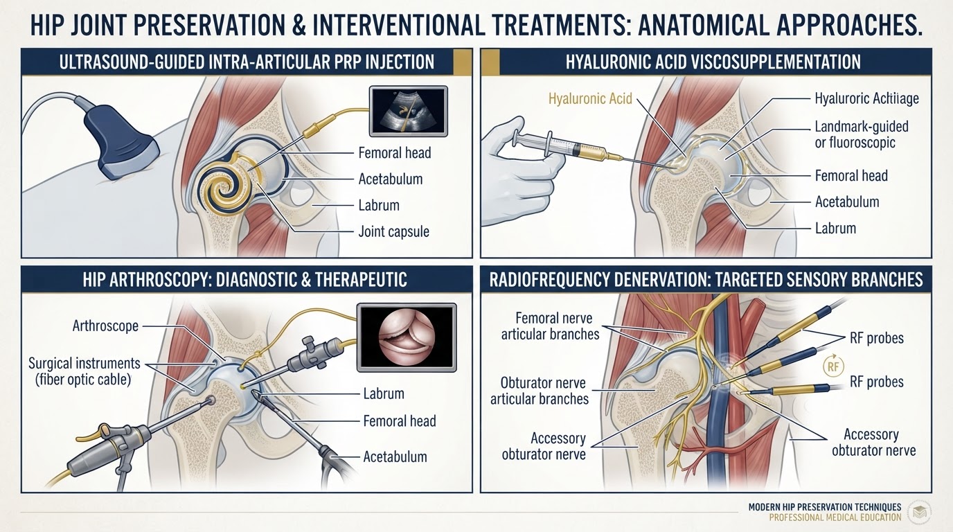
Platelet-rich plasma (PRP) and plasma rich in growth factors (PRGF) are autologous blood products prepared by centrifuging the patient's own blood to obtain a platelet concentrate that is injected into the joint. The rationale is that platelet-derived growth factors may modulate inflammation and influence joint biology. The evidence in hip osteoarthritis is more limited and more heterogeneous than in knee osteoarthritis. Some randomised controlled trials and systematic reviews suggest PRP may provide symptomatic improvement in selected patients, particularly with less advanced disease, with benefits reported at 3, 6 and 12 months in some studies; other trials find no clear advantage over hyaluronic acid or placebo. Comparisons are complicated by differences in PRP formulation (leukocyte-rich versus leukocyte-poor), single versus multiple injection protocols, and the severity of osteoarthritis studied. The 2026 VA/DoD guideline and relevant systematic reviews reflect this uncertainty. It is essential to be clear about what PRP is not: it is not a cartilage-regenerating treatment, and it does not reverse established osteoarthritis. No optimal PRP formulation, injection volume, protocol or number of sessions is established, and none is invented here. PRP is a symptomatic option to be considered individually, not a disease-modifying cure.
Hyaluronic acid injections
Hyaluronic acid (viscosupplementation) is injected into the hip joint to supplement synovial fluid, with the aim of improving lubrication and reducing pain. The evidence is conflicting. The AAOS 2023/2024 clinical practice guideline on management of osteoarthritis of the hip recommends against routine intra-articular hyaluronic acid injection, reflecting the body of evidence that does not reliably demonstrate a clinically important benefit. EUROVISCO 2026 recommendations provide a more nuanced position, supporting consideration of viscosupplementation in selected patients with appropriate indications, using image-guided injection. Randomised trials comparing hyaluronic acid with placebo or other injections show variable results, with some reporting short-term symptomatic benefit and others no clear advantage, and benefit appears more plausible in less advanced disease. Several points must be made honestly: hyaluronic acid is not a cartilage-regenerating treatment; different viscosupplementation products are not necessarily equivalent; and hip injections should be image-guided to ensure accurate intra-articular placement. The evidence does not support presenting hyaluronic acid as a proven disease-modifying therapy, and the conflicting recommendations between guideline bodies reflect genuine uncertainty.
Radiofrequency treatment for hip pain
Radiofrequency treatment of the hip targets the articular sensory branches that innervate the hip joint — principally the femoral articular branches, the obturator articular branches and, when relevant, the accessory obturator nerve. The aim is to interrupt pain signals from the joint by applying thermal (conventional), cooled or pulsed radiofrequency energy to these sensory branches. Conventional thermal radiofrequency creates a discrete lesion; cooled radiofrequency creates a larger, more spherical lesion that may better cover the variable anatomy of these branches; pulsed radiofrequency uses lower temperatures and is sometimes considered, with a different proposed mechanism. Patient selection typically involves diagnostic blocks of the relevant branches first, to confirm that the joint is the source of pain and that the targeted branches carry that pain signal. Published studies report pain and functional improvement in selected patients, with benefit that may last several months, though the evidence base is smaller and less mature than for knee genicular radiofrequency and results vary. It is critical to distinguish evidence from knee genicular radiofrequency from evidence specifically involving hip articular branches — the two are not interchangeable, and knee results should not be extrapolated to the hip. Radiofrequency is a pain-management intervention: it does not restore cartilage or correct mechanical degeneration. Its role is mainly in patients who are not surgical candidates, who wish to defer surgery, or in whom surgery is not appropriate, and it does not replace a properly indicated hip replacement.
Other image-guided injections and interventional options
Beyond PRP, hyaluronic acid and radiofrequency, other interventional options include intra-articular corticosteroid injections, which provide short-term anti-inflammatory pain relief and may be useful for flares (though repeated injections are not recommended as a long-term strategy), and periarticular injections for associated trochanteric pain syndrome or tendinopathies. All hip injections should be image-guided (ultrasound or fluoroscopy) to ensure accurate placement, as blind hip injections have a significant misplacement rate. These interventions are tools within a broader management plan, not a substitute for the core conservative measures or for surgery when it is indicated. Their role is to manage symptoms and support function while the underlying condition is monitored, and their use should be weighed against the evidence, the patient's response and the risk of simply delaying appropriate definitive treatment.
Hip arthroscopy: indications and limitations
Hip arthroscopy is a minimally invasive procedure that allows the surgeon to visualise and treat intra-articular pathology through small portals. It is important to state clearly what arthroscopy is not: it is not a treatment for established advanced hip osteoarthritis. Arthroscopy cannot replace lost cartilage or reverse a degenerative joint. Its role is in selected patients with early degeneration and a separate treatable intra-articular pathology — most commonly femoroacetabular impingement with labral tears and limited chondral damage — where correcting the underlying mechanical problem and addressing the labrum may relieve symptoms and potentially slow progression. Good candidates typically have preserved joint space, a treatable impingement morphology, a repairable labrum, minimal to moderate chondral damage, no significant subchondral bone change and no major osteophytes causing mechanical restriction. Joint-space measurement matters: a joint space of approximately 2 mm or less is associated with worse outcomes and a higher rate of conversion to total hip replacement, but this is a predictor among several, not an absolute universal rule — age, the extent of chondral damage, the presence of subchondral changes and the overall clinical picture all contribute. The Tönnis and Kellgren-Lawrence grades help frame this assessment. Counselling must be realistic: patients with significant degenerative change have higher rates of conversion to hip replacement after arthroscopy, and arthroscopy in advanced disease can worsen symptoms.
When should hip arthroscopy no longer be considered?
Hip arthroscopy should generally not be considered when osteoarthritis is established and advanced. Specific features that predict poor outcomes and argue against arthroscopy include severe joint-space narrowing (commonly cited as approximately 2 mm or less, but interpreted alongside other factors), high Tönnis or Kellgren-Lawrence grades, significant chondral damage (especially full-thickness cartilage loss), subchondral bone changes (oedema or cysts), large osteophytes causing mechanical restriction, older age with clear degenerative disease, and the absence of a treatable intra-articular lesion. In these situations arthroscopy is unlikely to help and may accelerate the need for hip replacement. The practical table below summarises how candidacy is assessed. The decision is always individualised: a borderline candidate with a clear, treatable mechanical problem and realistic expectations may still be considered by an experienced hip preservation surgeon, while a poor candidate should be directed toward conservative management or hip replacement rather than an operation that is unlikely to succeed. Hip arthroscopy candidacy — good candidate / borderline candidate / poor candidate (advanced osteoarthritis): * Good candidate: preserved joint space, treatable femoroacetabular impingement morphology, repairable labral tear, minimal chondral damage, no significant subchondral change, younger patient, realistic expectations. * Borderline candidate: moderate joint-space narrowing, some chondral damage, mixed degenerative and mechanical features — decision individualised by an experienced surgeon after detailed imaging. * Poor candidate / advanced osteoarthritis: joint space ≈2 mm or less, high Tönnis/Kellgren-Lawrence grade, full-thickness cartilage loss, subchondral changes, large osteophytes, older patient with established degenerative disease — arthroscopy not recommended; consider conservative management or hip replacement.
Joint-preserving procedures and selected alternatives
Beyond arthroscopy, joint-preserving surgery includes open or arthroscopic procedures to correct femoroacetabular impingement (cam, pincer or mixed), periacetabular osteotomy for hip dysplasia, and other realignment procedures in carefully selected younger patients. The goal is to correct a mechanical abnormality that is driving joint damage, ideally before significant osteoarthritis has developed. These procedures are most effective when performed early, in patients with preserved joint space and a correctable morphology, and they are not appropriate when the joint is already arthritic. Joint preservation is a philosophy of intervening to protect the native hip where possible, not a guarantee of avoiding replacement. For many patients with established osteoarthritis, the realistic choice is between ongoing conservative management and hip replacement, and presenting joint preservation as a universal alternative to replacement would be misleading.
When does conservative treatment stop being reasonable?
Conservative treatment does not have a fixed end point, but it stops being reasonable when it no longer provides meaningful benefit and the patient's quality of life is significantly affected. Indicators that conservative management is being exhausted include persistent pain despite appropriate non-operative measures, night pain and rest pain that disturb sleep, progressive walking limitation, loss of independence in daily activities, increasing reliance on analgesics with side effects, and repeated injections that provide only short-lived relief. Continuing to add ineffective treatments simply to defer surgery is not beneficial and can prolong suffering and disability. The ACR/AAHKS guidance on the optimal timing of elective hip arthroplasty supports moving to surgery when moderate-to-severe symptomatic osteoarthritis is present and non-operative treatment has failed, rather than persisting with treatments that are not working. There is no virtue in postponement for its own sake.
When should total hip replacement be recommended?
Total hip replacement is recommended when a patient has persistent, symptomatic moderate-to-severe hip osteoarthritis that has not responded adequately to appropriate conservative treatment, and the patient is medically fit for surgery. The decision integrates several factors: persistent pain (including night and rest pain), walking limitation and loss of independence, difficulty with everyday activities, progressive loss of range of motion, radiographic findings consistent with advanced osteoarthritis, failure or unsuitability of conservative and interventional treatment, the patient's expectations and preferences, and overall medical fitness. It is explicitly not based on an X-ray grade alone — a patient with severe radiographic change but good function may not need surgery, while a patient with moderate change but severe symptoms may. The ACR/AAHKS guideline on timing of elective arthroplasty emphasises that once moderate-to-severe symptomatic osteoarthritis is present and non-operative treatment has failed, delaying surgery to try additional ineffective treatments is not necessarily beneficial. The decision is made jointly between surgeon and patient through shared decision-making.
Can surgery be delayed safely?
In many patients, surgery can be delayed safely while conservative management is working and symptoms are tolerable — there is no obligation to operate the moment osteoarthritis is diagnosed, and many people never need a replacement. Delay is reasonable when it is purposeful: when conservative measures are providing meaningful benefit, when the patient is improving or stable, or when the patient prefers to wait. However, delay is not automatically safe or virtuous. Waiting becomes problematic when it means persisting with ineffective treatments, accepting increasing disability and pain, or losing conditioning and muscle strength that will make recovery harder. The decision to delay should be an active, considered one, reviewed regularly, not a default of inertia. For patients who are clearly candidates for hip replacement, indefinite postponement is not a neutral choice.
What happens if surgery is postponed for too long?
Postponing an indicated hip replacement for too long can have real consequences. These include prolonged unnecessary pain and disability, loss of independence, muscle wasting and deconditioning that make postoperative recovery slower and harder, deterioration of the joint that can make surgery more complex (for example, greater bone loss or more extensive deformity), and the development of comorbidities or decline in general health with age that increase surgical risk. Some studies suggest that very late presentation can be associated with somewhat worse functional outcomes, although hip replacement remains highly effective even in long-standing disease. The honest message is that there is a window in which surgery is most straightforward and recovery is easiest, and that waiting well beyond that window is not free — it carries its own costs in pain, function and operative complexity. This is why the decision is balanced, not simply 'delay as long as possible'.
Hip replacement approaches: anterolateral and direct superior approaches
When hip replacement is indicated, the surgical approach is one of several considerations. The anterolateral approach and the direct superior approach are muscle-sparing techniques that aim to minimise soft-tissue trauma and support early recovery; the direct anterior approach is another muscle-sparing option. Different approaches have different trade-offs in terms of exposure, dislocation risk, recovery speed and surgeon experience, and no single approach is universally superior — the best approach is the one your surgeon is most experienced with and that is appropriate for your anatomy and condition. The choice of approach does not change the fundamental decision of when to operate, and it should be discussed as part of the surgical plan rather than as a marketing point. Our dedicated article on anterior versus posterior hip replacement discusses surgical approaches in detail.
Recovery and expected outcomes after hip replacement
Total hip replacement is one of the most successful and cost-effective operations in orthopaedic surgery, with high rates of pain relief, functional improvement and patient satisfaction, and excellent long-term implant survival in registry data. Recovery is typically faster than after knee replacement: many patients walk on the day of or the day after surgery, are discharged within a few days, and return to most everyday activities within weeks, with continued improvement over several months. Full recovery and return to more demanding activities depend on the individual, the approach, the rehabilitation and the patient's general health. It is important not to promise a particular outcome or recovery time, because individual recovery varies. The expected outcome is substantial and durable pain relief and a major improvement in function and quality of life for the large majority of patients — but, as with any operation, results cannot be guaranteed for every individual.
Individualised treatment planning and conclusion
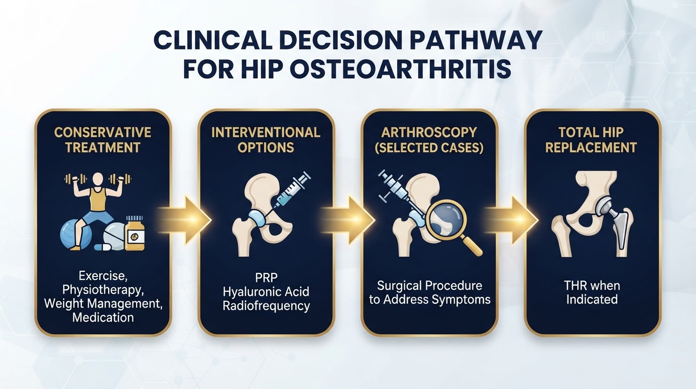
The management of hip osteoarthritis is not a single pathway but an individualised plan that evolves with the patient. The practical decision pathway below summarises how treatment progresses from early disease through to hip replacement. The key principles are: conservative management is the foundation and remains relevant throughout; interventional options (PRP, hyaluronic acid, radiofrequency) are symptomatic tools to be used selectively and honestly, not cures; hip arthroscopy is appropriate only in selected patients with treatable intra-articular pathology and limited degeneration; and hip replacement is recommended when moderate-to-severe symptomatic osteoarthritis has failed appropriate non-operative treatment, based on symptoms and function as much as on imaging. For residents and visitors across the Costa Blanca — Alicante, Torrevieja and Orihuela Costa — Dr Pablo Banchero offers individualised, evidence-based assessment and a frank second opinion to help you decide whether to preserve your hip or whether the time has come to replace it. Practical treatment algorithm: * EARLY HIP OSTEOARTHRITIS — Exercise, physiotherapy, activity management, appropriate medication. Consider selected image-guided interventions. Assess response and functional progress. * MILD–MODERATE HIP OSTEOARTHRITIS — Individualised conservative treatment. Consider PRP/PRGF or other interventions after discussing evidence limitations. Consider hip arthroscopy only when a separate treatable intra-articular pathology exists and joint degeneration is limited. Reassess symptoms and function. * ADVANCED HIP OSTEOARTHRITIS — Assess pain, disability, radiographs and quality of life. Discuss total hip replacement when conservative treatment is ineffective or unsuitable. Avoid repeated interventions that only delay appropriate surgery without meaningful benefit.
Preserve when you can, replace when you should
Hip osteoarthritis is managed across a spectrum from conservative care through interventional and joint-preserving options to hip replacement. PRP and hyaluronic acid may offer symptomatic relief in selected patients but do not regenerate cartilage or reverse advanced disease; radiofrequency is a pain-management tool, not a mechanical solution; hip arthroscopy is appropriate only for selected patients with treatable pathology and limited degeneration. Hip replacement is recommended when moderate-to-severe symptomatic osteoarthritis has failed appropriate non-operative treatment — based on symptoms and function, not an X-ray grade alone. On the Costa Blanca — Alicante, Torrevieja and Orihuela Costa — Dr Pablo Banchero provides evidence-based, individualised advice and second opinions on hip osteoarthritis and the timing of surgery.
References & scientific evidence
- American Academy of Orthopaedic Surgeons (AAOS). Management of Osteoarthritis of the Hip — Evidence-Based Clinical Practice Guideline.. AAOS Clinical Practice Guidelines, 2023.Clinical guidelineView source
- National Institute for Health and Care Excellence (NICE). Osteoarthritis in over 16s: diagnosis and management. NICE guideline NG226.. NICE Clinical Guidelines, 2022.Clinical guidelineView source
- American College of Rheumatology (ACR) / American Association of Hip and Knee Surgeons (AAHKS). Optimal Timing of Elective Hip or Knee Arthroplasty — Guideline.. Arthritis Care & Research / Journal of Arthroplasty, 2024.Clinical guidelineView source
- US Department of Veterans Affairs and Department of Defense (VA/DoD). VA/DoD Clinical Practice Guideline for the Non-Surgical Management of Hip and Knee Osteoarthritis.. VA/DoD Clinical Practice Guidelines, 2026.Clinical guidelineView source
- EUROVISCO. EUROVISCO recommendations for viscosupplementation of the hip joint.. EUROVISCO Consensus Recommendations, 2026.Clinical guidelineView source
- Díaz-Lledó A, Sánchez-Lázaro FJ, Alcorochel-Garrido P, et al.. Platelet-rich plasma in the treatment of hip osteoarthritis: a systematic review and meta-analysis.. Journal of Clinical Medicine, 2023.Systematic reviewDOI: 10.3390/jcm12062134View source
- Kuffner K, Capin JJ, Jelsing EJ, et al.. Intra-articular platelet-rich plasma injections for hip osteoarthritis: a randomized controlled trial.. Orthopaedic Journal of Sports Medicine, 2022.Randomised controlled trialDOI: 10.1177/23259671221107040View source
- Migliore A, Procopio S, Pignata A, et al.. Hyaluronic acid for hip osteoarthritis: a narrative review and practical considerations.. Therapeutic Advances in Musculoskeletal Disease, 2022.Observational studyDOI: 10.1177/1759720X221096540View source
- Sofat N, Kunupeddi P, Welton L, et al.. Comparative effectiveness of intra-articular hyaluronic acid and corticosteroid injections for hip osteoarthritis: a randomized controlled trial.. Rheumatology, 2023.Randomised controlled trialDOI: 10.1093/rheumatology/kead003View source
- Kapoor R, Liu AT, Darrith B, et al.. Radiofrequency denervation for chronic hip pain: a systematic review of articular branch interventions.. Pain Medicine, 2023.Systematic reviewDOI: 10.1093/pm/pnad045View source
- Karaman H, Kaya S, Tüfek A, et al.. Cooled radiofrequency ablation of the hip articular branches for chronic hip pain: clinical outcomes.. Pain Physician, 2022.Observational studyView source
- Domb BG, Martin TJ, Gu C, et al.. Predictors of clinical outcomes after hip arthroscopy: a 5-year prospective multicenter study.. American Journal of Sports Medicine, 2020.Observational studyDOI: 10.1177/0363546520909333View source
- Degen RM, Pan TJ, Chang B, et al.. Risk of total hip arthroplasty after hip arthroscopy in patients with pre-existing osteoarthritis.. Journal of Hip Preservation Surgery, 2018.Observational studyDOI: 10.1093/jhps/hny015View source
- Maldonado DR, Chen AW, Lall AC, et al.. Hip arthroscopy in patients with Tönnis grade 2 osteoarthritis: outcomes and conversion to total hip arthroplasty.. Journal of Arthroplasty, 2021.Observational studyDOI: 10.1016/j.arth.2021.02.038View source
- National Joint Registry (NJR) for England, Wales, Northern Ireland and the Isle of Man. National Joint Registry 21st Annual Report — Hip, Knee, Ankle, Elbow and Shoulder Arthroplasty: Outcomes and Survivorship.. National Joint Registry, 2024.Observational studyView source
Frequently asked questions

Dr. Pablo Banchero Lasalvia
Orthopaedic Surgeon · Traumatology Team
- Author:
- Dr. Pablo Banchero Lasalvia
- Medical reviewer:
- Dr. Pablo Banchero Lasalvia
- Published:
- 2 November 2026
- Medical review date:
- 2 November 2026
- ·
- 22 min read
Book Your Consultation via WhatsApp
Request a private, evidence-based orthopaedic assessment with Dr Pablo Banchero to discuss conservative care, PRP, hyaluronic acid, radiofrequency, hip arthroscopy or the timing of hip replacement on the Costa Blanca.
Official number: +34 602 99 22 22