
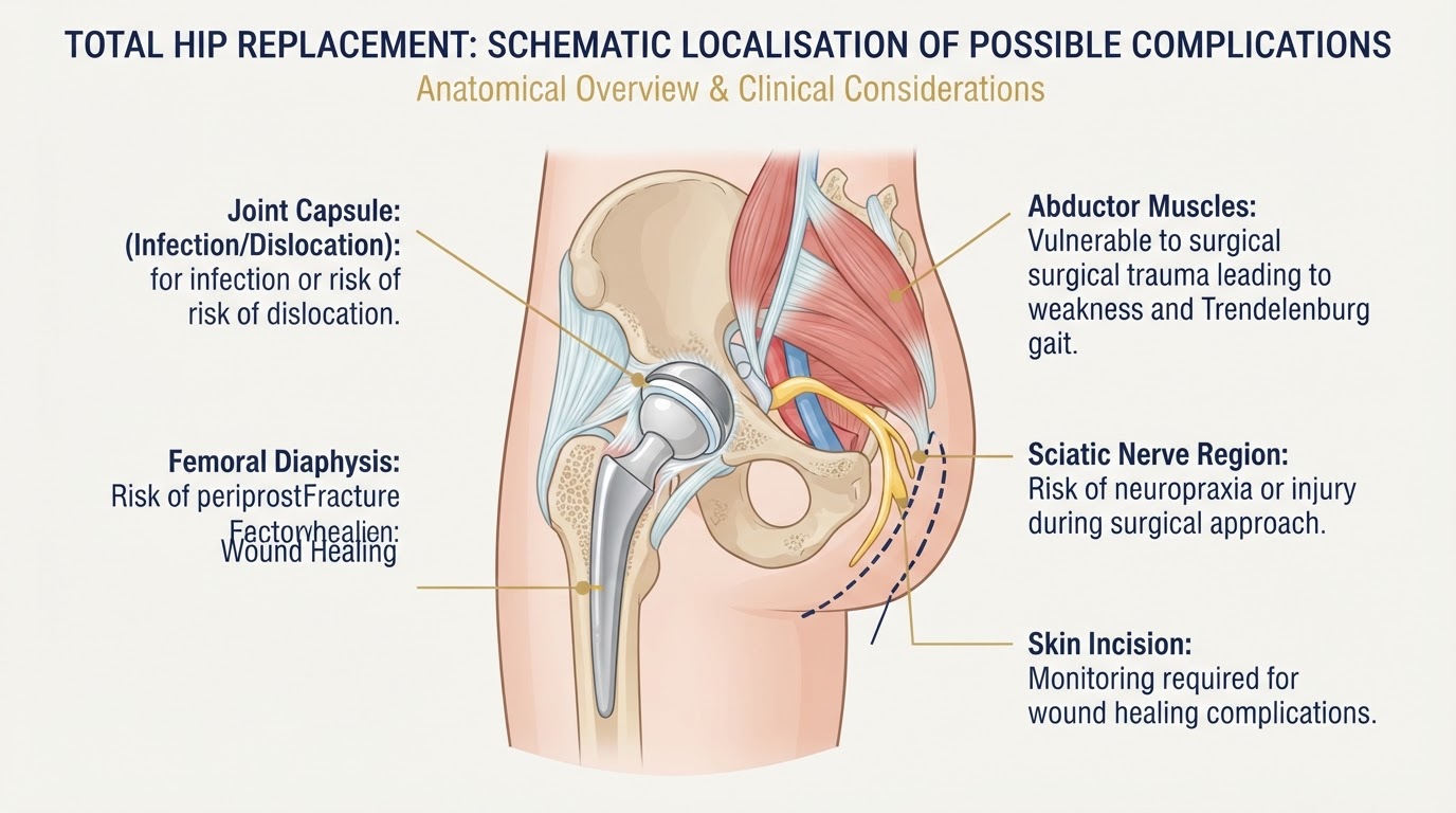
A total hip replacement can deliver excellent, long-lasting results, but — like every surgical procedure — it is never entirely free of risk. Understanding the possible complications, their warning signs and the measures that reduce them is an essential part of preparing for surgery. This article explains the main complications of hip replacement and how, at TraumatologyTeam, we approach risk reduction as a continuous process that begins before surgery and continues through the operation and into recovery. We do not promise the absence of complications or guaranteed outcomes; instead, we focus on identifying each patient's individual risk factors and establishing preventive measures adapted to their situation.
Understanding the Risks of Hip Replacement
Total hip replacement is one of the most effective and reliable operations in orthopaedic surgery, but no operation is risk-free. Complications can be grouped to help patients understand when they may occur and what they mean: • Intraoperative complications: events during the operation itself, such as a fracture around the implant, nerve or vessel injury, or anaesthetic issues. • Early postoperative complications: problems in the first days and weeks, including infection, bleeding or haematoma, blood clots, wound-healing difficulty and early dislocation. • Late complications: problems that appear months or years later, such as aseptic loosening, polyethylene wear, osteolysis, late dislocation, periprosthetic fracture or periprosthetic joint infection. • Implant-related problems: wear, breakage, instability or adverse local tissue reactions linked to the materials used. • Patient- or healing-related problems: delayed wound healing, persistent pain, stiffness or muscle weakness. Prevention is not a single act performed in the operating room. It is a continuous process that starts at the first consultation, continues through planning and surgery, and extends into structured follow-up.
How We Approach Risk Reduction at TraumatologyTeam
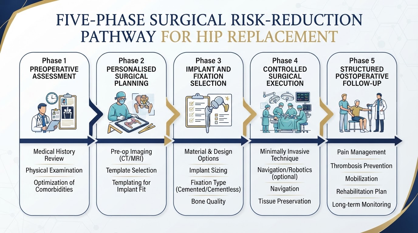
At TraumatologyTeam we structure risk reduction around five connected phases, each with specific preventive objectives: 1. Individual preoperative assessment — identifying medical, biological and functional risk factors before surgery. 2. Personalised surgical planning — analysing anatomy, leg length, offset and component orientation in advance. 3. Appropriate implant and fixation selection — matching the implant to bone quality, anatomy and activity rather than using the same solution for everyone. 4. Controlled surgical execution — careful soft-tissue handling, precise component positioning and intraoperative stability checks. 5. Structured postoperative follow-up — monitoring recovery, detecting warning signs early and guiding rehabilitation. This pathway does not eliminate complications — no clinical pathway can — but it allows potential problems to be anticipated and addressed before they become serious. The measures described in this article reflect established, evidence-based orthopaedic practice; where a specific protocol depends on individual circumstances, it is discussed with the patient rather than applied automatically.
Preoperative Assessment: Identifying Risk Before Surgery
A careful preoperative assessment is the foundation of prevention. We evaluate factors that can influence both surgical and healing risk: • Biological age and functional status. • Bone quality and osteoporosis. • Diabetes and glycaemic control. • Smoking, which impairs wound and bone healing. • Obesity and body composition. • Anaemia and nutritional status. • Cardiovascular and respiratory disease. • Previous thrombosis or clotting disorders. • Active infections (dental, urinary or skin), which should be treated first. • Medication, especially anticoagulants and immunosuppressants. • Previous hip surgery. • Fall risk and frailty. Identifying these factors does not create universal contraindications. It allows the patient to be optimised before surgery — for example improving glycaemic control, correcting anaemia, stopping smoking or adjusting medication — and allows the surgical plan to be adapted to the individual. We do not assign fixed risk percentages without a supporting source.
Preventing Periprosthetic Joint Infection
Periprosthetic joint infection (PJI) is one of the most serious complications of hip replacement. Infections may be superficial (limited to the wound) or deep (involving the implant), and are classified as acute or chronic depending on how soon they appear and the bacteria involved. Bacteria can reach the joint through contamination at the time of surgery or later through the bloodstream. Risk factors include poorly controlled diabetes, obesity, smoking, malnutrition, immunosuppression and active remote infections. Evidence-based preventive measures include: • Antibiotic prophylaxis according to protocol. • Skin preparation and antisepsis. • Aseptic surgical technique. • Glycaemic control where indicated. • Treatment of active infections before elective surgery. • Careful tissue handling and wound closure. • Operating-room environmental controls and protocol discipline. • Verification of instruments and components. NICE (NG157) and the AAOS guidance on periprosthetic joint infection support a layered, multimodal approach rather than relying on any single measure. No isolated measure eliminates the risk of infection; prevention depends on the combination of all of them.
Preventing Hip Dislocation
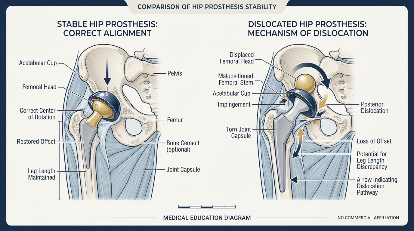
Dislocation occurs when the femoral head comes out of the acetabular cup. It is influenced by component orientation (acetabular inclination and anteversion, femoral version, combined anteversion), soft-tissue tension and offset, impingement, individual anatomy, previous surgery, neuromuscular disorders and spinopelvic pathology. Preventive measures include: • Preoperative planning of component position. • Restoration of biomechanics, offset and leg length. • Appropriate selection of head size and liner. • Intraoperative assessment of stability. • Selective use of dual mobility in patients at higher risk of dislocation. • Postoperative education adapted to the individual risk. As discussed in Article 18 (Direct Superior vs Anterolateral Hip Replacement) and Article 19 (Implant Selection, Fixation, Bearing Surfaces and Longevity), no single surgical approach eliminates the risk of dislocation. Stability depends on the combination of component position, soft-tissue balance and patient-specific factors.
Direct Superior and Anterolateral Approaches: Stability Considerations
The approaches used in our practice — the direct superior approach and the anterolateral approach — each have specific anatomical and stability considerations. The direct superior approach is a posterior-based, tissue-sparing technique usually performed in lateral decubitus. It works through a defined interval and allows management of the posterior capsule and short external rotators according to the specific technique, with stability assessed before closure. The anterolateral approach uses the interval between the tensor fasciae latae and the gluteus medius, and can be performed in lateral decubitus or supine depending on the variant and operating-room setup. It relates closely to the abductor mechanism, and muscle-sparing variants aim to preserve abductor function. We do not present these approaches as universally superior to others, and we do not extrapolate results from different approaches. Stability and recovery depend on component positioning, soft-tissue management and patient factors, not on the approach alone.
Leg Length Discrepancy
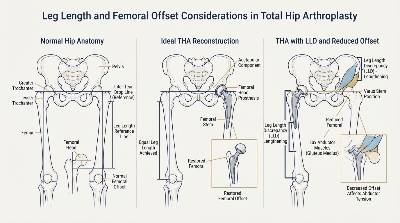
A difference in leg length after hip replacement may be real (an actual difference in bone length) or functional/perceived (related to offset, pelvic obliquity, muscle adaptation or pre-existing contractures). Restoring offset is as important as restoring length, because offset maintains abductor tension and joint stability. Preventive measures include: • Preoperative assessment of any existing discrepancy and pelvic obliquity. • Radiographic templating and planning. • Intraoperative comparison of anatomical references. • Restoration of offset and leg length. • Intraoperative checks balancing length and stability. Achieving perfectly identical leg length in every patient is not always possible and should not compromise stability or function. A small, planned difference is sometimes the safer choice to ensure a stable joint.
Preventing Periprosthetic Fractures
Periprosthetic fractures can occur during surgery (intraoperative) or afterwards (postoperative), and may affect the femur or, less commonly, the acetabulum. Risk factors include osteoporosis, older age, femoral morphology, the type of fixation used, previous surgery and falls. Preventive measures include: • Assessment of bone quality. • Individualised stem selection (cemented, cementless or hybrid according to bone). • Careful femoral preparation technique. • Controlled implant insertion to avoid excessive force. • Specific precautions in osteoporotic patients. • Patient education on fall prevention. These are preventive measures. When a fracture has already occurred, treatment options — fixation, revision of components or a combination — are different decisions taken according to the fracture pattern and implant stability.
Nerve and Vascular Injuries
Nerve injury is uncommon but possible. Structures at risk include the sciatic nerve, the femoral nerve and cutaneous nerves, with vascular injuries being rare. Associated factors include anatomy, previous surgery, retraction, leg lengthening and tension, and anatomical variations. Preventive measures rely on planning, careful exposure and controlled surgical execution: understanding the patient's anatomy, avoiding excessive retraction, limiting lengthening to what is biomechanically necessary, and recognising variations intraoperatively. Most nerve injuries are transient neuropraxia, but any new sensory or motor symptom after surgery should be reported and assessed.
Venous Thromboembolism: DVT and Pulmonary Embolism
Deep vein thrombosis (DVT) and pulmonary embolism (PE) are recognised risks after hip replacement. Risk factors include age, obesity, immobility, previous thrombosis and certain medical conditions. Warning signs include calf pain and swelling, chest pain, breathlessness or sudden shortness of breath. Preventive measures include: • Individual risk stratification (thrombotic and bleeding risk). • Pharmacological prophylaxis when indicated. • Early mobilisation. • Mechanical measures where appropriate. • Patient education on warning signs. We do not prescribe a universal anticoagulant or a fixed duration. Prophylaxis depends on the balance between thrombotic and bleeding risk and on the clinical protocol for each patient.
Wound Healing and Postoperative Haematoma
Wound problems include haematoma, bleeding, dehiscence (separation of the wound edges), delayed healing and secondary infection. They are more likely in patients with diabetes, obesity, smoking, malnutrition or those on anticoagulants. Good wound care, keeping the incision clean and dry, recognising warning signs early and avoiding premature strain all help. Increasing redness, spreading swelling, new discharge, worsening pain or fever should prompt contact with the surgical team.
Aseptic Loosening and Implant Failure
Aseptic loosening — loss of implant fixation without infection — is one of the most common late causes of failure. It is often driven by polyethylene wear particles that provoke osteolysis (bone resorption around the implant). Other late problems include component migration, mechanical failure and, with historical metal-on-metal bearings, adverse local tissue reactions. Modern highly cross-linked polyethylene and ceramic bearings have substantially reduced wear, but no bearing is immune to time and load. This is why long-term radiographic follow-up matters: detecting early osteolysis or loosening before symptoms become severe. This topic is covered in detail in Article 19.
Persistent Pain After Hip Replacement
Most patients have marked pain relief after hip replacement. When pain persists, the cause must be identified rather than assumed. Possible causes include: • Fixation problems or loosening. • Low-grade infection. • Iliopsoas tendinopathy (irritation of the psoas tendon against the cup). • Trochanteric pain or bursitis. • Biomechanical issues (offset or leg-length mismatch). • Referred lumbar pain. • Muscle injury or weakness. • Occult periprosthetic fracture. Persistent pain requires a structured assessment — history, examination, blood tests, imaging and sometimes aspiration — and should not be automatically attributed to the prosthesis without investigation.
Heterotopic Ossification
Heterotopic ossification is the formation of bone in soft tissues where it should not form after surgery. It can cause stiffness and, in severe cases, restricted movement. Risk factors include male sex, hypertrophic osteoarthritis, previous heterotopic ossification, ankylosing spondylitis and extensive surgical dissection. In selected higher-risk patients, prophylaxis with medication (such as non-steroidal anti-inflammatory drugs) or a single dose of radiotherapy may be considered when there is a specific indication. We do not recommend universal prophylaxis; it is reserved for patients with a clear increased risk.
Infection Warning Signs: When Should You Seek Help?
Contact the surgical team promptly — or attend urgent care — if you notice any of the following after hip replacement: • Fever or feeling generally unwell. • Progressive redness around the wound. • Wound discharge or leakage. • Increasing pain rather than the expected gradual improvement. • Abnormal swelling. • Wound dehiscence (edges opening). Early contact allows assessment before a superficial problem becomes a deep infection. Do not wait to see if it settles on its own.
Dislocation Warning Signs
A dislocated hip replacement usually causes sudden severe hip or groin pain, an apparent shortening or rotation of the leg, an inability to bear weight and an abnormal position of the limb that is difficult or impossible to move. A suspected dislocation requires urgent assessment and imaging. It should not be reduced at home. Emergency care ensures the hip is relocated safely, under appropriate control, and that the cause is reviewed to reduce the chance of recurrence.
Periprosthetic Fracture Warning Signs
After a fall or trauma, seek urgent assessment if you experience: • Severe pain around the hip or thigh. • Inability to bear weight. • Visible deformity. • Sudden loss of mobility. A periprosthetic fracture is a serious injury that needs prompt imaging and surgical assessment. Continuing to walk on a suspected fracture can worsen the damage.
How Are Complications Diagnosed?
Diagnosis depends on the suspected complication, but a structured workup commonly includes: • Clinical history and physical examination. • X-rays of the hip. • Blood tests, including inflammatory markers (ESR and CRP) when infection is suspected. • Joint aspiration with culture when infection is possible. • CT or other imaging when component position, fracture or loosening need detailed assessment. Not every test is needed in every case. The choice is guided by the clinical picture, and the diagnosis is built from the combination of findings rather than a single result.
What Happens If a Complication Occurs?
Treatment depends on the complication and the individual situation. General options include: • Conservative management when appropriate. • Closed or open reduction for dislocation. • Antibiotic treatment and surgery for selected infections, including debridement, antibiotics and implant retention (DAIR) in suitable patients. • Revision of one or both components for loosening, wear, instability or fracture. • Osteosynthesis (fixation) of periprosthetic fractures, sometimes combined with component revision. Each situation is different and should not be reduced to a single formula. The decision balances the type of complication, the implant status, the patient's general condition and their preferences.
Why Surgical Planning Matters
Careful planning is one of the most effective preventive tools available. Preoperative planning includes: • Radiological assessment of the hip and femur. • Femoral and acetabular anatomy. • Leg length and offset. • Femoral and acetabular version. • Bone quality. • Implant selection. • Surgical approach. • Planned final stability. Planning does not eliminate risk, but it allows potential problems to be identified and decisions adapted before the operation begins — rather than reacting to them during surgery.
Our Surgical Philosophy: Prevention Rather Than Reaction
Our approach is built on a few principles: • We do not treat every patient as if they were the same. • We select the implant according to anatomy and individual characteristics. • We plan the restoration of leg length, offset and stability. • We consider bone quality before choosing fixation. • We pay attention to soft-tissue preservation and handling. • We assess stability before finishing the operation. • We provide clear postoperative instructions. • We maintain clinical follow-up. This is a philosophy of professional responsibility and prevention. We do not claim that our results are better than those of other centres without a comparative audit to support it. What we can do is apply a disciplined, individualised process that addresses the details that influence safety and outcome.
Recovery and Patient Responsibilities
Patients play an active role in reducing risk during recovery: • Follow the postoperative instructions given. • Perform the mobilisation and exercises recommended. • Keep the wound clean and protected. • Avoid unauthorised activities during recovery. • Take prescribed prophylaxis as directed. • Attend follow-up appointments. • Report new symptoms promptly. Recovery is a partnership. Clear communication between patient and surgical team helps detect problems early and supports a safer return to function.
Questions to Ask Your Hip Surgeon
When planning hip replacement, consider asking: 1. What complications are most relevant in my case? 2. How do you assess my bone quality? 3. How will you plan implant positioning? 4. How do you reduce the risk of dislocation? 5. How do you assess and manage leg length? 6. What infection-prevention measures are used? 7. How is my risk of blood clots assessed? 8. What symptoms should make me contact the clinic? 9. What follow-up will I need? 10. Who should I contact if I have a concern after surgery?
Final Clinical Perspective
Complications of hip replacement cannot be eliminated completely, but many can be prevented or reduced through careful assessment, individualised planning, appropriate surgical technique and structured follow-up. The quality of a hip replacement is not measured only by implanting a prosthesis, but by taking the right decisions for each patient before, during and after the operation. Understanding the risks — and the measures that reduce them — allows patients to approach surgery with realistic expectations and to participate actively in their own recovery.
Prevention as a continuous, individualised process
At TraumatologyTeam, on the Costa Blanca — Alicante, Torrevieja, Orihuela Costa and La Zenia — we approach hip replacement with a philosophy of active prevention: individual preoperative assessment, personalised planning, implant selection matched to each patient, controlled surgical execution and structured follow-up. This does not eliminate complications, but it addresses the details that influence safety and outcome. If you are planning a hip replacement, a consultation with Dr Pablo Banchero allows your individual risks and options to be discussed in detail.
References & scientific evidence
- NICE Guideline NG157. Joint replacement (primary): hip, knee and shoulder. Clinical guideline.. National Institute for Health and Care Excellence, 2020.Clinical guidelineView source
- AAOS Clinical Practice Guideline. Diagnosis and Prevention of Periprosthetic Joint Infections of the Hip and Knee.. American Academy of Orthopaedic Surgeons, 2019.Clinical guidelineView source
- AAOS/AAHKS Clinical Practice Guideline. Management of Patients Undergoing Elective Hip or Knee Arthroplasty: Dental Interventions and the Prevention of Periprosthetic Joint Infection.. American Academy of Orthopaedic Surgeons, 2024.Clinical guidelineView source
- Parvizi J, Tan TL, Goswami K, et al.. The 2018 Definition of Periprosthetic Hip and Knee Infection: An Evidence-Based and Validated Criteria.. The Journal of Arthroplasty, 2018.Observational studyDOI: 10.1016/j.arth.2018.02.078View source
- Bozic KJ, Kurtz SM, Lau E, et al.. The epidemiology of revision total hip arthroplasty in the United States.. The Journal of Bone and Joint Surgery (American), 2009.Observational studyDOI: 10.2106/JBJS.H.00155View source
- Kurtz SM, Lau E, Watson H, Schmier JK, Parvizi J.. Economic burden of periprosthetic joint infection in the United States.. Journal of Arthroplasty, 2012.Observational studyDOI: 10.1016/j.arth.2012.02.022View source
- Lewinnek GE, Lewis JL, Tarr R, Compere CL, Zimmerman JR.. Dislocations after total hip-replacement arthroplasties.. Journal of Bone and Joint Surgery (American), 1978.Observational studyPMID: 670295View source
- Vigdorchik JM, Sharma AK, Elbuluk AM, et al.. Spinopelvic dysplasia and hypermobility increase dislocation risk in total hip arthroplasty: a functional planning protocol.. The Bone & Joint Journal, 2021.Observational studyDOI: 10.1302/0301-620X.103B7.BJJ-2020-2391.R1View source
- Bourassa-Moreau M, Lavigne S, Pelet S, et al.. Heterotopic ossification after total hip arthroplasty: a systematic review.. Journal of Arthroplasty, 2020.Systematic reviewDOI: 10.1016/j.arth.2020.04.025View source
- Garcia DC, Darrith B, Davies CM, Bohl DD, Bhandari M, Della Valle CJ.. Periprosthetic Joint Infection Following Hip and Knee Arthroplasty: A Review of Diagnosis and Management.. JAMA Surgery, 2024.Systematic reviewDOI: 10.1001/jamasurg.2024.0153View source
- Gulhane S, Lanting B, Teeter M, McCalden R, Vasarhelyi E.. Leg length discrepancy and offset following total hip arthroplasty: a review of the literature and current practice.. HIP International, 2021.Systematic reviewDOI: 10.1177/1120700020974197View source
- Cichocki AR, Law TY, Rosas S, et al.. Periprosthetic femur fractures after total hip arthroplasty: a systematic review of risk factors.. Orthopedic Reviews, 2019.Systematic reviewDOI: 10.4081/or.2019.7772View source
- Falck-Ytter Y, Francis CW, Johanson NA, et al.. Prevention of VTE in Orthopedic Surgery Patients: Antithrombotic Therapy and Prevention of Thrombosis, 9th ed.. Chest (ACCP Guidelines), 2012.Clinical guidelineDOI: 10.1378/chest.11-2398View source
- National Joint Registry (NJR) for England, Wales, Northern Ireland and the Isle of Man. National Joint Registry 21st Annual Report — Hip, Knee, Ankle, Elbow and Shoulder Arthroplasty: Outcomes and Survivorship.. National Joint Registry, 2024.Observational studyView source
- Australian Orthopaedic Association National Joint Replacement Registry (AOANJRR). Hip, Knee & Shoulder Arthroplasty: Annual Report 2024.. AOA NJRR, 2024.Observational studyView source
- Della Valle CJ, Parvizi J, Bauer TW, et al.. American Academy of Orthopaedic Surgeons clinical practice guideline on: the diagnosis of periprosthetic joint infection of the hip and knee.. Journal of Bone and Joint Surgery (American), 2011.Clinical guidelineDOI: 10.2106/JBJS.K.00558View source
- Abdel MP, Watts CD, Wuthrich PS, Berry DJ, Hanssen AD.. Outcomes and complications of revision total hip arthroplasty in the setting of periprosthetic femoral fracture.. The Bone & Joint Journal, 2017.Observational studyDOI: 10.1302/0301-620X.99B6.BJJ-2016-0776.R1View source
- Schwarzkopf R, Baghoolizadeh M, Long WJ, Della Valle CJ.. Dislocation Following Total Hip Arthroplasty: A Comprehensive Review.. Journal of the American Academy of Orthopaedic Surgeons, 2024.Systematic reviewDOI: 10.5435/JAAOS-D-23-00570View source
Frequently asked questions

Dr. Pablo Banchero Lasalvia
Orthopaedic Surgeon · Traumatology Team
- Author:
- Dr. Pablo Banchero Lasalvia
- Medical reviewer:
- Dr. Pablo Banchero Lasalvia
- Published:
- 2 November 2026
- Medical review date:
- 2 November 2026
- ·
- 25 min read
Discuss Your Individual Risks and Surgical Options via WhatsApp
Planning a hip replacement? Request a private orthopaedic consultation with Dr Pablo Banchero to discuss individual risks, surgical planning and the preventive measures that apply to your case.
Official number: +34 602 99 22 22