
One of the most common and most important questions in hip care is simply: how do I know when it is time for a hip replacement? The honest answer is that there is no single test, X-ray measurement or age that decides it. The decision is made when the combination of persistent symptoms, loss of function, radiographic evidence and the patient's own goals points clearly toward surgery, and when reasonable conservative treatment has either failed or is unsuitable. This article explains what happens in advanced hip osteoarthritis, which symptoms suggest surgery may be appropriate, when conservative treatment has reached its limits, whether there is an ideal age, how implants and surgical approaches are chosen, and what to expect during recovery. It is written for residents and visitors on the Costa Blanca — including patients in Alicante, Torrevieja and Orihuela Costa — and for international patients seeking an evidence-based second opinion. The central message is balanced: surgery should not be rushed, but it should also not be postponed indefinitely when pain, immobility and loss of independence are clearly progressing and conservative options are no longer helping.
Introduction: how do you know when it is time for hip replacement?
Hip replacement is one of the most successful and reliable operations in orthopaedic surgery, but deciding the right moment is a clinical judgement, not a formula. The decision is never based on an X-ray grade alone. Some patients have severe radiographic osteoarthritis but manage well and do not need surgery; others have moderate changes on imaging but disabling pain and loss of function for whom surgery is clearly appropriate. The right time is when persistent symptoms, functional limitation and the failure of reasonable conservative treatment align with the patient's goals and overall medical fitness — discussed openly between surgeon and patient. This article walks through that decision so you can approach it with realistic expectations, neither rushing toward an operation you do not yet need, nor postponing one that is clearly indicated.
What happens in advanced hip osteoarthritis?
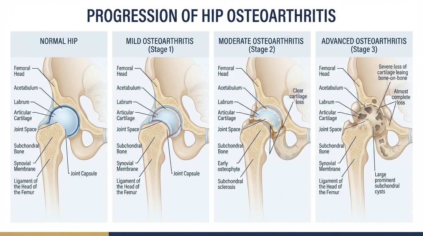
Hip osteoarthritis is the progressive loss of the articular cartilage that covers the femoral head and the acetabulum. As cartilage thins, the joint space narrows on X-ray, the underlying bone becomes sclerotic and may form subchondral cysts, and osteophytes (bone spurs) develop at the joint margins. In advanced disease the cartilage may be worn through to bone, producing mechanical bone-on-bone contact. This drives pain that is increasingly present at rest and at night, stiffness that limits movement, and progressive loss of function. Understanding this progression matters because it explains why conservative treatments that modulate inflammation or symptoms may help earlier but cannot correct the structural problem once the joint is severely degenerated.
Symptoms that may indicate the need for surgery
Several symptoms, in combination, suggest that surgery may be appropriate. Persistent groin or thigh pain that limits daily life is central. Pain at rest or at night that disturbs sleep is particularly significant, because it indicates that the joint is symptomatic even when unloaded. Increasing difficulty walking, climbing stairs, putting on shoes and socks, or rising from a chair reflects loss of function. Progressive stiffness and a feeling that the hip is 'giving way' or unreliable are also relevant. No single symptom is decisive, but the pattern of persistent, worsening pain and functional loss despite reasonable non-operative care is the typical picture that prompts surgical consideration.
Pain during walking, rest and at night
The character and timing of pain matter. Early osteoarthritis pain is usually activity-related — it comes on with walking and eases with rest. As the joint degenerates, pain becomes more persistent, present during rest, and then at night, often disturbing sleep. Night pain is a particularly important symptom because it signals that the joint is symptomatic even when unloaded, and it often has a major impact on quality of life. When night or rest pain becomes persistent and is not relieved by reasonable conservative measures, it is one of the stronger indicators that surgery may be appropriate. The decision is still individualised, but persistent rest and night pain should not be dismissed as something to simply live with.
Loss of mobility and limitations in daily activities
Function is as important as pain. Many patients adapt gradually to a shrinking world — walking less, avoiding stairs, giving up activities, needing help with dressing or footwear, and losing independence. This functional decline is sometimes noticed more by family than by the patient. When osteoarthritis is limiting walking distance, making stairs difficult, interfering with personal care, or causing loss of independence, these are important indicators that surgery may improve quality of life. The aim of hip replacement is not only to reduce pain but to restore function and independence, and functional limitation is a legitimate and central reason to consider it.
Clinical examination and radiographic assessment
A clinical examination assesses range of motion, gait, leg length, muscle strength and the pattern of pain. Radiographs (X-rays) confirm the presence and severity of osteoarthritis — joint-space narrowing, sclerosis, cysts and osteophytes — and help rule out other causes of hip pain. Grading systems such as Kellgren-Lawrence and Tönnis describe the radiographic severity. Both the examination and the imaging contribute to the decision, but neither alone determines it. The examination confirms that the symptoms come from the hip, and the imaging confirms the structural disease — but the decision to operate integrates these with the patient's symptoms, function and goals.
Why X-rays alone do not determine the need for surgery
This is a crucial point that is often misunderstood. There is no single radiographic threshold that determines the need for a hip replacement. The correlation between X-ray appearance and symptoms is imperfect: some patients with severe radiographic osteoarthritis have manageable symptoms and do not need surgery, while others with moderate changes have disabling pain that is clearly helped by surgery. The decision is based on the combination of symptoms, function, imaging and shared decision-making — not on an X-ray grade alone. A 'bad X-ray' is not by itself a reason to operate, and a 'not-so-bad X-ray' is not a reason to withhold surgery from a patient who is clearly suffering and losing function.
When conservative treatment has reached its limits
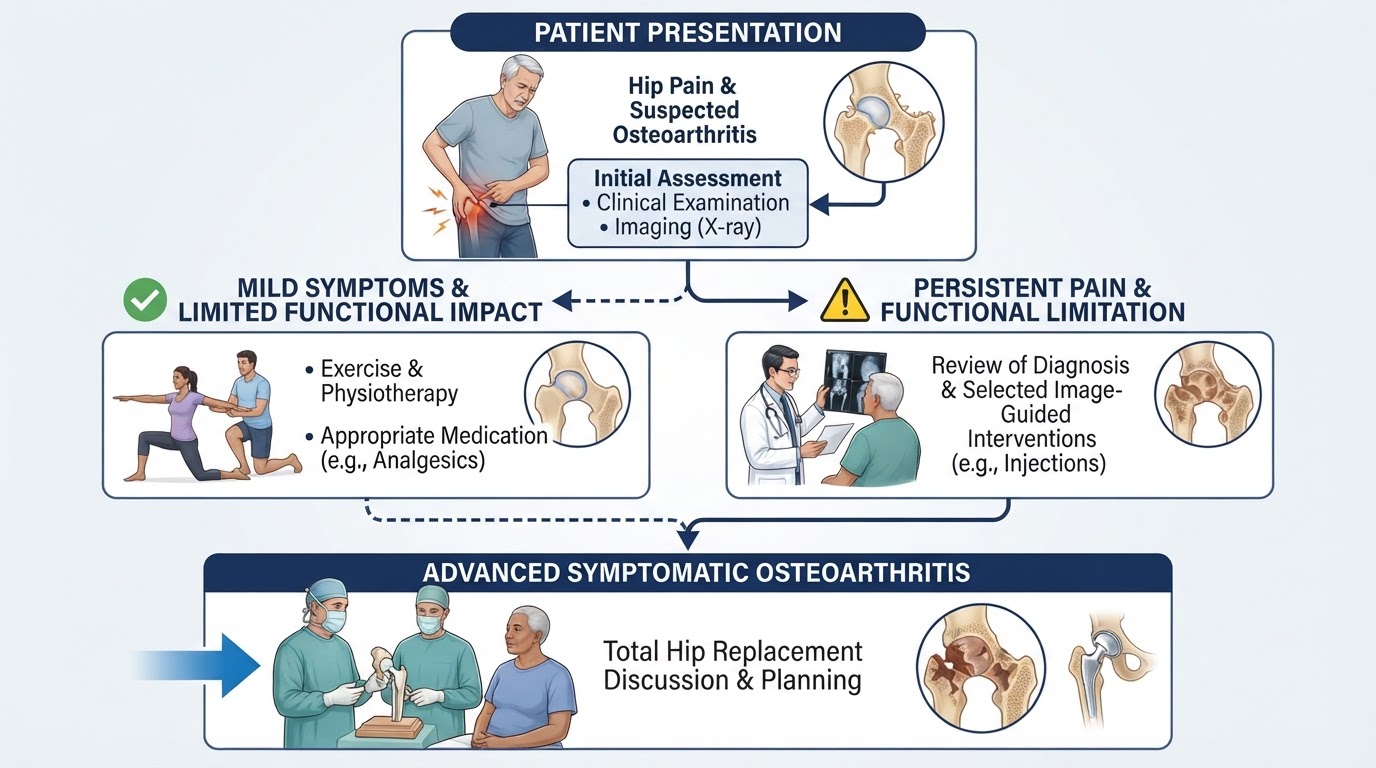
Conservative treatment does not have a fixed end point, but it stops being reasonable when it no longer provides meaningful benefit and quality of life is significantly affected. Indicators that conservative management is being exhausted include persistent pain despite appropriate non-operative measures, night and rest pain that disturb sleep, progressive walking limitation, loss of independence in daily activities, increasing reliance on analgesics with side effects, and repeated injections that provide only short-lived relief. Continuing to add ineffective treatments simply to defer surgery is not beneficial and can prolong suffering and disability. The ACR/AAHKS guidance on the optimal timing of elective hip arthroplasty supports moving to surgery when moderate-to-severe symptomatic osteoarthritis is present and non-operative treatment has failed, rather than persisting with treatments that are not working. There is no virtue in postponement for its own sake.
Physiotherapy, exercise and lifestyle management
Before surgery is considered, conservative management is the foundation. Therapeutic exercise and physiotherapy maintain range of motion and muscle strength, which protect the joint and improve function. Weight management, where appropriate, reduces load on the hip. Activity modification — pacing, avoiding aggravating activities, using walking aids when needed — helps patients remain functional. Appropriate analgesics and anti-inflammatory medication, when suitable, control symptoms. These measures are genuinely effective for many patients and should be pursued. The point at which surgery is considered is when these measures have been given a reasonable trial and are no longer providing adequate relief, not when they have been skipped.
PRP/PRGF and hyaluronic acid: when they may still be considered
Intra-articular platelet-rich plasma (PRP/PRGF) and hyaluronic acid are sometimes offered for hip osteoarthritis. The evidence is limited and conflicting: PRP may provide symptomatic improvement in selected patients, particularly with less advanced disease, but does not regenerate lost cartilage or reverse advanced osteoarthritis; hyaluronic acid recommendations are not uniform across professional bodies (AAOS recommends against routine use, EUROVISCO supports selected use), and different products are not necessarily equivalent. These injections may still be considered in early-to-moderate disease in selected patients after an honest discussion of the evidence and its limitations. They are not a reason to defer a clearly indicated replacement in advanced symptomatic disease, and they should not be repeated indefinitely when they are not helping.
Radiofrequency and other pain-management interventions
Radiofrequency treatment of the hip's sensory articular branches is a pain-management intervention that may reduce pain for a period in selected patients with refractory hip pain, particularly those who are not surgical candidates or wish to defer surgery after informed discussion. It does not treat the arthritis, regenerate cartilage or restore joint space, and knee genicular evidence cannot be extrapolated to the hip. It may have a role in carefully selected patients, but it is not a substitute for a hip replacement when the joint is severely degenerated and surgery is indicated. Like injections, it should not be used to postpone a clearly indicated operation indefinitely.
Hip arthroscopy: when preservation surgery is no longer appropriate
Hip arthroscopy is not a treatment for established advanced hip osteoarthritis. It may be considered in selected patients with early degeneration and a separate treatable intra-articular problem (such as femoroacetabular impingement with labral pathology) where the joint space is preserved. Once osteoarthritis is advanced — severe joint-space narrowing, diffuse cartilage loss, osteophytes and stiffness — arthroscopy has a poor prognosis and a higher likelihood of early conversion to total hip replacement. In advanced symptomatic disease, hip arthroscopy is generally not appropriate, and total hip replacement is the more suitable option. The boundary between preservation surgery and replacement is based on the degree of degeneration, the treatable pathology and realistic expectations, not on a wish to avoid a prosthesis.
When should total hip replacement be recommended?
Total hip replacement is recommended when a patient has persistent, symptomatic moderate-to-severe hip osteoarthritis that has not responded adequately to appropriate conservative treatment, and the patient is medically fit for surgery. The decision integrates several factors: persistent pain (including night and rest pain), walking limitation and loss of independence, difficulty with everyday activities, progressive loss of range of motion, radiographic findings consistent with advanced osteoarthritis, failure or unsuitability of conservative and interventional treatment, the patient's expectations and preferences, and overall medical fitness. It is explicitly not based on an X-ray grade alone. The ACR/AAHKS guideline on the timing of elective arthroplasty emphasises that once moderate-to-severe symptomatic osteoarthritis is present and non-operative treatment has failed, delaying surgery to try additional ineffective treatments is not necessarily beneficial. The decision is made jointly between surgeon and patient through shared decision-making.
Is there an ideal age for hip replacement?
There is no universally ideal age for hip replacement. The decision is based on symptoms, function, imaging and fitness, not on a number. Historically, some patients were told to wait until they were 'old enough', but modern implants and techniques have changed this. Younger patients may have a greater lifetime probability of eventually requiring a revision, which is a real consideration to discuss, but it is not a reason to deny surgery to a younger patient who is clearly suffering and losing function. Older patients may have additional medical and functional considerations, but age alone is not a contraindication. Biological health, comorbidities, bone quality, activity level and expectations matter more than chronological age. The right question is not 'am I the right age?' but 'is my hip problem severe enough, and am I well enough, for surgery to improve my life?'
Hip replacement in younger and older patients
In younger patients, the main consideration is that a prosthesis may need to last many decades, and the lifetime probability of revision is higher than in an older patient. Registry data and long-term survival studies provide reassurance about modern implant durability, but a younger patient should understand the possibility of future revision and the importance of activity choices that protect the joint. In older patients, the considerations are different: comorbidities, bone quality, frailty and surgical fitness are more relevant, and the balance of benefit and risk is individualised. In both groups, the decision is based on the overall clinical picture, not on age alone. A fit older patient can do very well, and a younger patient with severe symptomatic disease should not be made to wait indefinitely.
What happens if surgery is postponed?
Postponing surgery is reasonable when conservative treatment is working and symptoms are manageable. But postponement is not always harmless. When a clearly indicated replacement is delayed for too long, the patient may endure prolonged pain, worsening stiffness, progressive muscle weakness and loss of conditioning, declining mobility and independence, and sometimes deterioration in general health that can make later surgery more difficult. There is also evidence that operating on a more deteriorated joint can make recovery harder. This does not mean surgery should be rushed, but it means that deferring an indicated operation indefinitely — particularly to try repeated ineffective injections — is not a neutral choice. The ACR/AAHKS guidance supports moving to surgery when the criteria are met rather than persisting with treatments that are not working.
Implant selection and fixation: cemented versus cementless
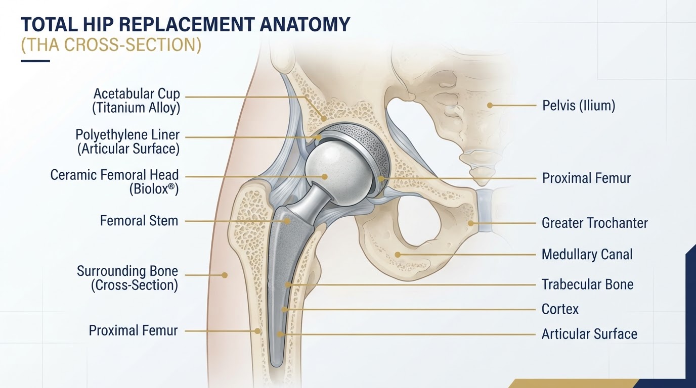
A total hip replacement has two main components: the acetabular cup, which is fixed into the pelvis, and the femoral stem, which is inserted into the femur, topped by a femoral head that articulates with a liner inside the cup. Fixation can be cemented (using bone cement), cementless (the component is designed for bone to grow onto its surface), or hybrid (one component cemented, the other cementless). Bearing surfaces vary — ceramic or metal femoral heads, and highly cross-linked polyethylene or ceramic liners — and each has different wear characteristics. No single implant design or fixation method is universally superior; the choice depends on patient anatomy, bone quality, age, activity level and surgical judgement. These decisions are individualised and discussed with the patient, not presented as one option being objectively best.
Surgical approaches: anterolateral and direct superior approaches
There are several surgical approaches to the hip, each with anatomical principles, potential advantages and limitations. The anterolateral approach may be performed in lateral or supine positioning depending on the technique; it accesses the hip through an interval that respects the abductor mechanism as far as possible, and is a well-established approach with reliable exposure. The direct superior approach is usually performed in lateral decubitus and is designed to be muscle-sparing, with a limited surgical exposure; it has a learning curve and is suited to selected patients. Neither approach is universally superior, and the choice depends on the surgeon's experience, the patient's anatomy and individual factors. The direct anterior approach is not the main technique addressed by this clinical programme and is not the focus here. The honest position is that a well-executed operation through any appropriate approach, with well-placed components, can achieve excellent results.
What to expect during recovery
Recovery after hip replacement is individual and depends on the approach, the patient's fitness and the surgical detail. In general, early mobilisation is encouraged, often on the day of surgery or the next, with walking aids used as needed. Physiotherapy guides the progression of movement, strengthening and gait. Pain is managed with a multimodal approach. Most patients return to many daily activities within weeks, and driving, work and sport are resumed progressively according to recovery and the surgeon's guidance. Long-term function is generally very good for the large majority of patients. No fixed recovery date can be guaranteed, because rehabilitation depends on the procedures performed and the patient's condition, but the overall trajectory is toward restored mobility and reduced pain.
Conclusion: making an individualised decision
The right time for hip replacement is not defined by an X-ray grade or an age, but by the combination of persistent symptoms, functional limitation, radiographic evidence and the patient's goals, once reasonable conservative treatment has been given a fair trial. Surgery should not be rushed, but it should also not be postponed indefinitely when pain, immobility and loss of independence are clearly progressing and conservative options are no longer helping. Implant and approach choices are individualised, and recovery is generally very rewarding for the large majority of patients. For residents and visitors across the Costa Blanca — Alicante, Torrevieja and Orihuela Costa — Dr Pablo Banchero offers an individualised, evidence-based assessment and an honest second opinion on whether conservative care, joint preservation or hip replacement is the right path for your hip.
An individualised, evidence-based decision
Hip replacement is recommended when persistent, symptomatic moderate-to-severe osteoarthritis has not responded to appropriate conservative treatment — based on symptoms, function, imaging and shared decision-making, not on an X-ray grade alone. There is no ideal age; biological health, comorbidities, bone quality and expectations matter more. Surgery should not be rushed, but postponing a clearly indicated operation to try repeated ineffective treatments is not necessarily beneficial. Implant fixation and surgical approach are individualised, and recovery is generally very rewarding. On the Costa Blanca — Alicante, Torrevieja and Orihuela Costa — Dr Pablo Banchero provides evidence-based assessment and second opinions to help you decide the right time for hip replacement.
References & scientific evidence
- National Institute for Health and Care Excellence (NICE). Osteoarthritis in over 16s: diagnosis and management. NICE guideline NG226.. NICE Clinical Guidelines, 2022.Clinical guidelineView source
- National Institute for Health and Care Excellence (NICE). Joint replacement (primary): hip, knee and shoulder. NICE guideline NG157.. NICE Clinical Guidelines, 2020.Clinical guidelineView source
- American Academy of Orthopaedic Surgeons (AAOS). Management of Osteoarthritis of the Hip — Evidence-Based Clinical Practice Guideline.. AAOS Clinical Practice Guidelines, 2023.Clinical guidelineView source
- American College of Rheumatology (ACR) / American Association of Hip and Knee Surgeons (AAHKS). Optimal Timing of Elective Hip or Knee Arthroplasty — Guideline.. Arthritis Care & Research / Journal of Arthroplasty, 2024.Clinical guidelineView source
- National Joint Registry (NJR) for England, Wales, Northern Ireland and the Isle of Man. National Joint Registry 21st Annual Report — Hip, Knee, Ankle, Elbow and Shoulder Arthroplasty: Outcomes and Survivorship.. National Joint Registry, 2024.Observational studyView source
- Australian Orthopaedic Association National Joint Replacement Registry (AOANJRR). Hip, Knee & Shoulder Arthroplasty: Annual Report.. AOA NJRR, 2024.Observational studyView source
- Evans JT, Evans JP, Walker RW, Blom AW, Whitehouse MR, Sayers A.. How long does a hip replacement last? A systematic review and meta-analysis of case series and national registry reports.. The Lancet, 2019.Meta-analysisDOI: 10.1016/S0140-6736(18)31665-9View source
- Bayliss LE, Culliford D, Monk AP, et al.. The effect of patient age at intervention on risk of implant revision after total hip replacement: population-based cohort study.. BMJ, 2017.Observational studyDOI: 10.1136/bmj.j1411View source
- Higgins BT, Barlow DR, Heagerty NE, Lin TJ.. Anterior vs. posterior approach for total hip arthroplasty: a systematic review and meta-analysis.. Journal of Arthroplasty, 2015.Meta-analysisDOI: 10.1016/j.arth.2014.12.009View source
- Miller LE, Gondusky JS, Kamath AF, Boettner F, Wright TM, Bhattacharyya S.. Influencing risk factors for instability in total hip arthroplasty: a systematic review and meta-analysis.. Orthopaedic Journal of Sports Medicine, 2020.Meta-analysisDOI: 10.1177/2325967120951834View source
- Petis S, Howard JL, Lanting BL, Vasarhelyi EM.. Surgical approach in primary total hip arthroplasty: anatomy, technique and clinical outcomes.. HIP International, 2015.Systematic reviewDOI: 10.5301/hipint.5000262View source
- Tsikandylakis I, Mohaddes M, Cnudde P, Eskelinen A, Kärrholm J, Rolfson O.. Cementless compared with cemented total hip arthroplasty: an analysis of 134,651 patients from the Swedish Hip Arthroplasty Register.. Journal of Bone and Joint Surgery (Am), 2018.Observational studyDOI: 10.2106/JBJS.17.00934View source
- Degen RM, Pan TJ, Chang B, et al.. Risk of total hip arthroplasty after hip arthroscopy in patients with pre-existing osteoarthritis.. Journal of Hip Preservation Surgery, 2018.Observational studyDOI: 10.1093/jhps/hny015View source
- Maldonado DR, Chen AW, Lall AC, et al.. Hip arthroscopy in patients with Tönnis grade 2 osteoarthritis: outcomes and conversion to total hip arthroplasty.. Journal of Arthroplasty, 2021.Observational studyDOI: 10.1016/j.arth.2021.02.038View source
- U.S. Department of Veterans Affairs and Department of Defense (VA/DoD). VA/DoD Clinical Practice Guideline for the Non-Surgical Management of Hip and Knee Osteoarthritis.. VA/DoD Clinical Practice Guidelines, 2026.Clinical guidelineView source
Frequently asked questions

Dr. Pablo Banchero Lasalvia
Orthopaedic Surgeon · Traumatology Team
- Author:
- Dr. Pablo Banchero Lasalvia
- Medical reviewer:
- Dr. Pablo Banchero Lasalvia
- Published:
- 2 November 2026
- Medical review date:
- 2 November 2026
- ·
- 20 min read
Book Your Consultation via WhatsApp
Request a private, evidence-based orthopaedic assessment with Dr Pablo Banchero to discuss persistent hip pain, whether conservative treatment remains appropriate, the timing of hip replacement, or a second opinion before surgery on the Costa Blanca.
Official number: +34 602 99 22 22