
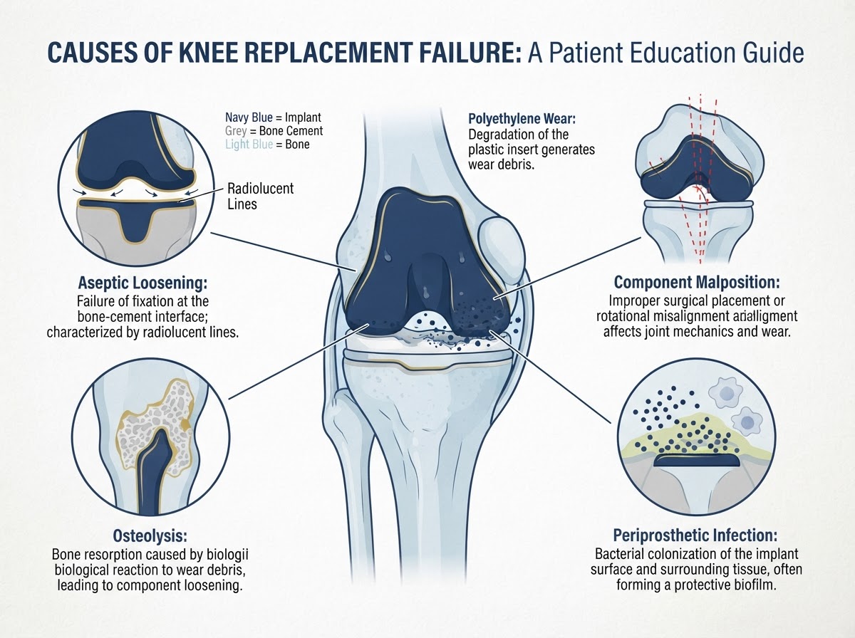
Most knee replacements relieve pain and restore function for many years, but a small proportion of implants stop working as they should. When that happens, it is rarely a single dramatic event: the more common picture is persistent or recurrent pain, new swelling, a feeling that the knee is giving way, or a gradual loss of movement. The challenge is that these symptoms overlap with ordinary recovery and with problems that have nothing to do with the prosthesis. This article explains what knee replacement failure actually means, how the symptoms differ from normal postoperative healing, how your surgeon investigates a painful knee replacement, and what treatment options exist — from conservative care through to revision surgery. It is written for residents and visitors on the Costa Blanca — including patients in Alicante, Torrevieja and Orihuela Costa — and for international patients seeking an English-speaking second opinion about a previous knee replacement.
Introduction: what does it mean when a knee replacement fails?
A knee replacement is considered to have 'failed' when it no longer delivers the pain relief and function that was expected, or when a problem with the implant or the joint means further treatment is needed. Failure is not a single diagnosis — it is an umbrella term that covers several distinct causes, each with its own pattern of symptoms, investigation and treatment. The most frequent reasons a knee replacement is revisited are aseptic loosening (the implant gradually loses its fixation to bone without infection), periprosthetic joint infection, polyethylene wear with osteolysis, instability, component malposition, patellofemoral problems and — less commonly — periprosthetic fracture or nerve problems. Importantly, a painful knee replacement is not automatically a failed one: many patients with persistent discomfort in the first months after surgery are simply still recovering, and many painful knees settle with time and rehabilitation. Failure implies a specific, identifiable cause that the workup is designed to find.
Difference between a painful knee replacement and a mechanically failed implant
Distinguishing a 'painful knee replacement' from a 'mechanically failed implant' is the central diagnostic question. A painful knee replacement is a symptom: the knee hurts, but the cause may be the implant, the soft tissues, referred pain from elsewhere, or the natural course of recovery. A mechanically failed implant is a specific diagnosis: the prosthesis itself is loose, worn, malpositioned, infected or unstable, and that mechanical problem is the source of the symptoms. The two are not the same. A patient can have a well-fixed, well-positioned, non-infected implant and still have pain — from stiffness, tendon irritation, hip or spine pathology, complex regional pain, or incomplete rehabilitation. Equally, an implant can show minor radiographic changes that are clinically silent. This is why a painful knee after replacement is not treated as a failure by default; it is investigated systematically until a cause is identified, and only then is a treatment plan — conservative, medical or surgical — built around that cause.
Common symptoms
The symptoms that prompt assessment of a knee replacement are usually familiar to patients who have lived with the joint for months or years. They include: * Persistent or recurrent pain: pain that does not continue to improve, or that returns after a pain-free interval. Pain on standing or weight-bearing is particularly relevant, as is aching that worsens with activity. * Swelling or recurrent effusions: a knee that repeatedly fills with fluid, feels warm or looks puffy, especially if this is new after a long stable period. * Instability or giving way: a sensation that the knee will buckle, particularly on stairs, turning or uneven ground. This may indicate ligament imbalance or component malposition rather than simple weakness. * Reduced range of motion: a loss of flexion or extension that is new, or stiffness that persists or progresses beyond the expected rehabilitation window. * Difficulty walking or climbing stairs: a functional decline — needing a stick or frame again, or avoiding activities that were previously manageable. * Systemic features when infection is present: fever, night pain, wound discharge or feeling generally unwell, though many infections present more subtly. None of these symptoms is, on its own, proof of failure. Their significance depends on timing: early postoperative symptoms are often part of recovery, whereas new symptoms after a long stable interval are more concerning and warrant assessment.
Aseptic loosening
Aseptic loosening is the gradual loss of fixation between an implant component and the surrounding bone or cement, occurring without infection. Over time, repetitive loading can disrupt the bone–cement or bone–implant interface, producing a radiolucent line (a dark gap on X-ray) and eventually allowing the component to migrate or tilt. Typical features are progressive, activity-related aching rather than acute pain, a sensation that the knee is 'shifting', and — on radiographs — widening radiolucent lines at the bone–cement interface or measurable migration of the component compared with earlier films. The mechanism is partly mechanical (repeated stress at the interface) and partly biological: microscopic wear particles from the bearing surface provoke a local inflammatory response that resorbs bone (osteolysis), which in turn accelerates loosening. Aseptic loosening is one of the commonest single reasons for revision knee replacement in the long term in registry data. Not every radiolucent line means the implant must be revised: the decision weighs the severity of loosening, the symptoms, the rate of change and the bone stock available for reconstruction.
Periprosthetic joint infection
Periprosthetic joint infection (PJI) is infection involving the prosthetic components and the joint space. It is one of the most important diagnoses to consider, because the consequences of treating a presumed aseptic failure when infection is present are serious. Infections are generally described by timing and severity: * Acute infection: presenting in the first weeks after surgery, often with wound redness, discharge, fever and increasing pain. These may follow wound-healing problems or early contamination. * Chronic (late) infection: presenting months or years later, sometimes after a long symptom-free interval. Symptoms may be subtle — new aching, night pain, recurrent effusions or a gradual decline in function — and can be triggered by bacteria reaching the bloodstream from distant sources (dental procedures, skin or urinary infections) and seeding the implant. * Acute haematogenous infection: a sudden, florid presentation in a previously well joint, often with marked pain, swelling and systemic upset. Because chronic infection can mimic aseptic loosening, and because revising an unrecognised infected knee as if it were aseptic carries a high risk of failure, infection must be investigated and reasonably excluded before any presumed aseptic revision. The validated international definition of PJI (the ICM criteria, reflected in the AAOS Clinical Practice Guideline on periprosthetic joint infections) combines clinical, laboratory, synovial and microbiological findings rather than relying on a single test. Treatment depends on timing and severity, the organism and the patient, and may range from antibiotic suppression through debridement with retention or exchange of modular components, to one- or two-stage revision with antibiotic-loaded cement.
Polyethylene wear and osteolysis
The bearing surface of most knee replacements is a polyethylene (plastic) insert that sits between the metal femoral and tibial components. Over millions of cycles of walking, this surface slowly wears, producing fine particles. These particles can provoke a biological response in the surrounding bone — osteolysis — in which macrophages resorb bone around the implant. Osteolysis may be painless early on and is often first detected on routine follow-up radiographs as cystic areas around the components. As wear progresses, the insert can thin, become uneven or, in advanced cases, fracture or dissociate, leading to instability, maltracking or rapid loosening. Polyethylene wear is therefore a long-term, gradual process rather than a sudden event, and it interacts with loosening: wear particles drive osteolysis, osteolysis undermines fixation, and loosening follows. The rate depends on the bearing material (modern highly cross-linked polyethylenes wear more slowly than older conventional polyethylene), patient activity, alignment and surgical technique. Detecting wear early — before structural damage is severe — is one of the goals of scheduled radiographic follow-up.
Instability and ligament imbalance
A knee replacement relies on balanced soft tissues as well as on the implant. Instability after knee replacement can take several forms: a sense of the knee giving way or buckling, recurrent effusions (a 'wet' knee is often an unstable one), hyperextension, or symptomatic mediolateral or anteroposterior (flexion) laxity. Causes include ligament imbalance or asymmetry at the time of the original surgery, progressive attenuation of the collateral ligaments, component rotation or malposition that alters the kinematics, and — in posterior-stabilised designs — failure of the cam-and-post mechanism. Symptomatic instability tends to present with giving-way on stairs, turning or uneven surfaces, rather than constant pain. It is distinguished from ordinary early postoperative weakness by persistence beyond the rehabilitation period and by a mechanical quality to the symptoms. Management depends on the cause and severity: a structured strengthening programme and bracing may help mild cases, while symptomatic instability that persists or is mechanically significant generally requires assessment for revision — which may involve balancing the soft tissues, exchanging the polyethylene insert, or revising one or more components.
Malalignment, component positioning and rotational problems
The way the components are positioned influences how the knee moves and how the loads are distributed. Component malalignment or malrotation can produce abnormal kinematics even when the components are well fixed: the patella may track poorly, the flexion gap may be asymmetric, and edge loading can accelerate wear. Rotational malposition of the femoral or tibial component in particular is associated with patellar maltracking, stiffness and pain. Symptoms may include anterior knee pain, a sense that the knee 'does not move naturally', stiffness in certain ranges, and asymmetric wear on follow-up imaging. Plain radiographs may suggest alignment problems, but computed tomography (CT) is the investigation of choice when component rotation or subtle malposition needs to be assessed in detail. As with loosening, not all malposition needs revision: the decision is based on whether the malalignment is the cause of the symptoms, whether it is accelerating wear or instability, and whether correcting it will reliably improve the knee without creating new problems.
Patellofemoral problems and patellar component complications
The kneecap (patella) and its articulation with the front of the femur (the trochlea) are a common focus of symptoms after knee replacement. Problems include: * Anterior knee pain: pain over the front of the knee, which may relate to the patellar component, soft-tissue tension, patellar overload or impingement. * Patellar maltracking: the kneecap does not glide centrally through its groove, often related to component rotation or lateral soft-tissue tightness, producing a catching or subluxation sensation. * Patellar component loosening or failure: in resurfaced patellae, the button can loosen, wear or dissociate, sometimes producing a palpable clunk or mechanical symptoms. * Patellar clunk syndrome: fibrous tissue at the superior pole catching in extension, producing a painful snap; this is usually managed conservatively or with arthroscopic release when persistent. Not all anterior discomfort after knee replacement is a patellar problem — early quadriceps weakness, swelling and rehabilitation soreness are common and settle — but persistent, localised anterior pain with mechanical features warrants assessment. Treatment ranges from physiotherapy and activity modification through to patellar resurfacing or component revision in selected cases.
Periprosthetic fractures
A periprosthetic fracture is a break in the femur or tibia immediately around the knee replacement. It is uncommon after primary surgery and more often seen with fragile bone, in revision procedures, after a fall, or where stress-risers from previous fixation devices or bone defects concentrate load. Risk is increased by osteoporosis, rheumatoid arthritis, long-term steroid use, stiffness requiring forceful manipulation, and component loosening that weakens the adjacent bone. The presentation is usually clear — new, severe pain and inability to bear weight after an injury — but a subtle or minimally displaced fracture can occasionally be missed on a single radiograph and emerge on comparison with previous films or CT. Treatment depends on the fracture pattern, the bone quality and, crucially, on whether the implant is still well fixed: a fracture around a stable component may be managed with internal fixation, while a fracture around a loose component usually requires revision with a long-stemmed prosthesis that bypasses and stabilises the fracture. Prevention respects bone health, falls precautions and surgical technique.
Painful knee replacement with no obvious radiographic abnormality
A particularly challenging group of patients have a painful knee replacement in which standard radiographs show no obvious loosening, wear, malposition or fracture. A normal-looking X-ray does not exclude every cause of pain. Several possibilities remain and are actively considered: a low-grade or chronic periprosthetic infection (which can be radiographically silent), referred pain from the hip or spine (hip arthritis or lumbar nerve-root compression can refer to the knee), soft-tissue causes (tendinopathy, impingement, complex regional pain), inadequate rehabilitation or stiffness, and — in a small minority — pain with no identifiable structural cause. This is exactly the setting in which the workup is extended: comparison with previous films, blood markers to screen for infection, and when indicated joint aspiration, CT to assess rotation, or nuclear medicine studies. The goal is not to label the knee as 'unexplained' but to methodically exclude the treatable causes before concluding that no structural problem is present. In many such patients a cause is found; in others, conservative management and reassurance are appropriate rather than surgery.
Diagnostic assessment
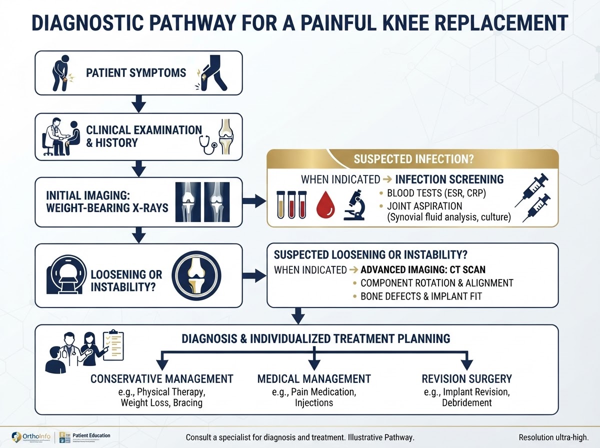
The investigation of a painful or suspected failed knee replacement is systematic and individualised. It is not a fixed protocol applied to every patient. The pathway, illustrated in the diagnostic diagram below, typically progresses through several stages: * Clinical history and examination: timing and pattern of the pain, change from the postoperative baseline, presence of instability or stiffness, wound history, systemic features, and the functional impact. Examination assesses the wound, alignment, range of motion, ligament stability, gait and the patellofemoral joint. * Weight-bearing radiographs: current weight-bearing anteroposterior and lateral views, a patellar/sunrise view, and long-leg films when alignment is in question. The single most useful investigation is comparison with previous imaging — migration, widening radiolucencies and new wear are seen by change over time. * Blood tests when infection is suspected: C-reactive protein (CRP) and erythrocyte sedimentation rate (ESR) as screening tests. These are sensitive but not specific: they help decide whether further infection workup is needed, and a normal result does not completely exclude infection in every case. * Joint aspiration and synovial analysis when indicated: when infection is suspected on screening, aspiration of synovial fluid for cell count (particularly the white blood cell count and differential), culture and, in some centres, leucocyte esterase or alpha-def Wassermann-type markers. Aspiration is a selective, targeted test, not a routine one. * CT for component rotation and bone defects: when malposition, rotational problems or bone loss need detailed assessment that radiographs cannot provide. CT is also useful for surgical planning before revision. * Nuclear medicine studies in selected cases: in difficult or equivocal situations, additional studies may be used selectively. They are not routine. The aim is to move from symptoms and examination to a working diagnosis — aseptic loosening, infection, instability, malposition, wear, referred pain, or a combination — and then to build an individualised treatment plan rather than a generic pathway.
How doctors distinguish infection from aseptic loosening
Distinguishing infection from aseptic loosening is the pivotal step, because the treatments are very different and treating an infected joint as if it were aseptic leads to recurrent failure. The validated approach, reflected in the international consensus definition and the AAOS Clinical Practice Guideline on periprosthetic joint infections, combines several lines of evidence rather than relying on a single test: * Clinical pattern: a sinus tract communicating with the joint is diagnostic of infection; acute onset with systemic features is suggestive of infection, while slow progression over years favours aseptic loosening. But chronic infection can be indolent and mimic loosening. * Serum inflammatory markers (CRP, ESR): used to screen for infection. They are not definitive standalone tests: normal values can occur in low-grade infection, and elevated values can follow recent surgery or other inflammation. Abnormal results prompt further workup. * Synovial fluid analysis: white cell count and differential, culture, and sometimes leucocyte esterase. A positive culture with a compatible organism is strong evidence; the cell-count thresholds used in the consensus definition are applied in context, not in isolation. * Imaging: radiographs may show periosteal reaction, rapidly progressive loosening or osteolysis that raises suspicion; nuclear medicine studies can help in selected cases. * Intraoperative findings at revision: frozen-section histology and multiple cultures from different sites, interpreted together, are part of the definitive workup when revision is undertaken. The point is that no single number — not the CRP, not a cell count, not an X-ray — makes the diagnosis in isolation. The combination does, and the judgement of an experienced surgeon remains central.
Treatment options
Treatment is built around the cause, not the symptoms alone. Not every painful or even mechanically failed knee requires revision surgery, and revision is not always the whole answer. The spectrum of options includes: * Conservative treatment when appropriate: for stiffness, soft-tissue causes or referred pain, a structured physiotherapy programme, activity modification, analgesia, weight management and, when relevant, treatment of the hip or spine. * Medical treatment of infection: in selected patients unfit for surgery, or as a bridge, long-term suppressive antibiotics may be used, though they do not eradicate established deep infection. * Treatment of infection with surgery: debridement, antibiotics and implant retention (DAIR) with exchange of modular components in early acuteute infection; one-stage or two-stage revision for chronic infection, with antibiotic-loaded cement and a dedicated interval spacer in two-stage protocols. * Isolated component or polyethylene exchange: in selected cases of isolated wear, maltracking or instability with well-fixed components, exchange of the insert or revision of a single component may be appropriate — but this is a careful, case-specific decision, not a default. * Full revision knee replacement: when one or more components are loose, malpositioned or infected, the failed components are removed, bone defects are reconstructed, and new components are implanted, often with stems, augments or cones. The choice depends on the diagnosis, the bone stock, the soft tissues, the patient's general health and expectations, and the surgeon's experience. There is no 'one size fits all' pathway.
What happens during revision knee replacement surgery?
Revision knee replacement is, in general, a larger and more complex operation than the primary procedure, and the recovery is correspondingly more demanding. The operation involves several stages: careful exposure of the joint (often through the previous scar, but occasionally requiring a more extensile approach), controlled removal of the failed components while preserving as much bone and soft tissue as possible, thorough debridement and, when infection is present, sampling and exchange of all foreign material; assessment and reconstruction of bone defects using bone graft, augments, sleeves or cones; and implantation of revision components, frequently with intramedullary stems to gain fixation in the femoral or tibial canal and to bypass defects. When infection is the indication, the strategy is usually either a one-stage revision (single operation) or a two-stage revision (removal and antibiotic spacer first, then reimplantation after a course of antibiotics), chosen on the basis of the organism, the soft tissues and the patient. The operation takes longer than a primary replacement, the hospital stay is usually longer, and the postoperative course is more variable. Because each revision is tailored to the individual knee, the technical plan is discussed and consented specifically rather than generalised.
Expected recovery and rehabilitation after revision
Recovery after revision knee replacement is more variable and often slower than after a primary replacement, and it depends heavily on the reason for revision, the bone reconstruction required, the presence of infection and the patient's general health. In general, early mobilisation and a structured rehabilitation programme are encouraged when clinically appropriate, but some situations — for example, a two-stage infection revision with a spacer in place, or reconstruction of a large bone defect — require protected weight-bearing or specific restrictions for a period. Physiotherapy focuses on restoring range of motion, quadriceps and hip strength, balance and gait, and is progressed individually rather than to a fixed timetable. It is not appropriate to give a single recovery timeline that applies to every revision: some patients return to most activities within a few months, while others with complex reconstructions follow a longer, phased plan. The practical message is that rehabilitation after revision is more bespoke than after a first replacement, and goals — for mobility, pain and function — should be set with your surgeon and physiotherapist on the basis of the specific operation performed.
When should a patient seek urgent medical attention?
Some problems related to a knee replacement should not wait for a routine appointment. Seek urgent assessment for: * Acute, severe pain and inability to bear weight after a fall or injury (possible periprosthetic fracture). * Spreading wound redness, increasing discharge, opening of the wound, or fever and chills (possible acute infection). * A sudden, marked change in the knee — new instability, a feeling that the knee will give way, or a sudden loss of movement — particularly after an injury. * Systemic symptoms such as persistent fever, night sweats or feeling increasingly unwell, which may indicate haematogenous infection seeding the implant. * Significant new calf pain, one-sided leg swelling, chest pain or shortness of breath (possible thromboembolism), which require emergency assessment. In contrast, a gradual ache that has been present for some time, mild swelling after activity, or the normal ebb and flow of recovery are reasons to book a planned review rather than to attend urgently. When in doubt, contact your surgical team: the safe approach is to be assessed promptly rather than to wait.
Questions to ask an orthopaedic surgeon
A second opinion about a previous knee replacement is most useful when you can give the surgeon a clear account of the problem. Useful questions include: * Based on my symptoms and imaging, which compartments or components are involved, and is there evidence of loosening, wear, malposition or instability? * Has infection been properly considered and excluded, and what tests were used? * Could my pain be coming from elsewhere — my hip or spine — rather than from the knee replacement itself? * If revision is being considered, what are the expected benefits, the limitations and the realistic recovery? * What is your experience with revision knee replacement, and how many do you perform? * What are the alternatives to revision in my case, and what happens if we wait? * If I have had previous surgery elsewhere, can you access my records and imaging? These questions help align the assessment with your individual situation rather than with a generic protocol, and they help you weigh the benefits and risks of further surgery.
Conclusion and consultation CTA
A knee replacement that stops working as expected is a problem that deserves a careful, systematic assessment rather than an assumption. Pain does not automatically mean the implant is loose; a normal-looking X-ray does not exclude every cause; and infection must be considered and excluded before any presumed aseptic revision. The right pathway moves from symptoms and examination through targeted imaging and laboratory tests to an individualised diagnosis, and then to a treatment plan that may be conservative, medical or surgical — and, when revision is needed, is tailored to the specific cause and to your knee. For residents and visitors across the Costa Blanca — Alicante, Torrevieja and Orihuela Costa — Dr Pablo Banchero offers individualised, evidence-based assessment of painful or failing knee replacements, whether you are seeking a diagnosis for the first time or a second opinion about a previous operation.
A careful assessment, not a hurried conclusion
Knee replacement failure is not a single diagnosis but a set of identifiable causes — loosening, infection, wear, instability, malposition, patellar problems, fracture or, sometimes, a cause outside the knee. The right approach is a systematic workup that distinguishes a painful knee from a mechanically failed implant, excludes infection, identifies the cause, and matches the treatment to that cause and to you as an individual. On the Costa Blanca — Alicante, Torrevieja and Orihuela Costa — Dr Pablo Banchero provides individualised orthopaedic assessment and revision expertise.
References & scientific evidence
- American Academy of Orthopaedic Surgeons (AAOS). Diagnosis and Prevention of Periprosthetic Joint Infections of the Hip and Knee — Evidence-Based Clinical Practice Guideline.. AAOS Clinical Practice Guidelines, 2019.Clinical guidelineView source
- American Academy of Orthopaedic Surgeons (AAOS). Surgical Management of Osteoarthritis of the Knee — Evidence-Based Clinical Practice Guideline (SMOAK2).. AAOS Clinical Practice Guidelines, 2022.Clinical guidelineView source
- Parvizi J, Tan TL, Goswami AK, Shea M, Comhey C, Kamath AF, et al.. The 2018 Definition of Periprosthetic Hip and Knee Infection: An Evidence-Based and Validated Criteria.. Journal of Arthroplasty, 2018.Observational studyDOI: 10.1016/j.arth.2018.02.058PMID: 29661564View source
- National Institute for Health and Care Excellence (NICE). Joint replacement (primary): hip, knee and shoulder. NICE guideline NG157.. NICE Clinical Guidelines, 2020.Clinical guidelineView source
- National Institute for Health and Care Excellence (NICE). Reducing risk of venous thromboembolism (deep vein thrombosis and pulmonary embolism) in patients going to hospital. NICE guideline NG89.. NICE Clinical Guidelines, 2018.Clinical guidelineView source
- Zimmerli W, Trampuz A, Ochsner PE. Prosthetic-joint infections.. New England Journal of Medicine, 2004.Systematic reviewDOI: 10.1056/NEJMra040168View source
- Evans JT, Walker RW, Evans JP, Blom AW, Sayers A, Whitehouse MR. How long does a knee replacement last? A systematic review and meta-analysis of case series and national registry reports.. The Lancet, 2019.Meta-analysisDOI: 10.1016/S0140-6736(18)32531-5View source
- National Joint Registry (NJR) for England, Wales, Northern Ireland and the Isle of Man. National Joint Registry 21st Annual Report — Hip, Knee, Ankle, Elbow and Shoulder Arthroplasty: Outcomes and Survivorship.. National Joint Registry, 2024.Observational studyView source
- Australian Orthopaedic Association National Joint Replacement Registry (AOANJRR). Hip, Knee & Shoulder Arthroplasty Annual Report 2024.. AOA NJRR, 2024.Observational studyView source
- Swedish Knee Arthroplasty Register (SKAR). The Swedish Knee Arthroplasty Register — Annual Report.. Swedish Knee Arthroplasty Register, 2023.Observational studyView source
- Riaz O, Aqil A, Husain A, Chishti I, Hill JP, Nicholl A. Periprosthetic fractures around total knee arthroplasty — a review.. Annals of Medicine and Surgery, 2021.Systematic reviewDOI: 10.1016/j.amsu.2021.102598View source
Frequently asked questions

Dr. Pablo Banchero Lasalvia
Orthopaedic Surgeon · Traumatology Team
- Author:
- Dr. Pablo Banchero Lasalvia
- Medical reviewer:
- Dr. Pablo Banchero Lasalvia
- Published:
- 2 November 2026
- Medical review date:
- 2 November 2026
- ·
- 20 min read
Book Your Consultation via WhatsApp
Request a private orthopaedic assessment with Dr Pablo Banchero for persistent pain after knee replacement, suspected loosening or implant failure, or an expert second opinion before revision surgery on the Costa Blanca.
Official number: +34 602 99 22 22