
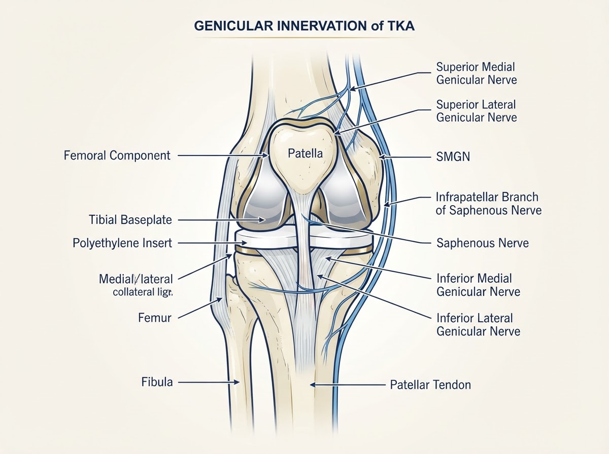
Most people feel substantially better after a knee replacement, but a meaningful minority continue to have pain longer than expected. For some, this is simply the slower end of normal recovery; for others, the pain points to a specific, treatable cause that the implant itself may or may not be responsible for. The difficulty is that persistent pain after knee replacement overlaps with ordinary healing, with problems outside the knee, and with several distinct diagnoses — mechanical loosening, low-grade infection, stiffness, neuropathic pain, tendinopathy and referred pain from the hip or spine — each needing a different response. This article explains why a knee can still hurt after replacement, how your surgeon works through the differential diagnosis, and the full range of treatment options, from targeted rehabilitation and genicular nerve radiofrequency through to revision surgery. It is written for residents and visitors on the Costa Blanca — including patients in Alicante, Torrevieja and Orihuela Costa — and for international patients seeking an English-speaking second opinion about a painful knee replacement.
Introduction: why can a knee still hurt after replacement?
A knee replacement is one of the most effective operations in orthopaedics for advanced osteoarthritis, but 'effective' does not mean 'pain-free for everyone immediately and permanently'. A proportion of patients continue to experience pain beyond the expected recovery window, and a smaller group describe pain that persists for months or years. The reasons are varied: some are part of a slow but normal recovery, some relate to the implant (loosening, wear, malposition, instability), some to infection, some to the soft tissues and nerves around the knee, and some to problems elsewhere — the hip or the lumbar spine — that refer pain to the knee. The central message of this article is that persistent pain is a symptom that deserves a systematic assessment, not a diagnosis in itself. The goal of that assessment is to identify the actual cause, because the right treatment — rehabilitation, medication, nerve intervention, biological therapy or revision surgery — depends entirely on what is actually driving the pain.
Expected postoperative discomfort versus persistent chronic pain
It is important to separate expected postoperative discomfort from persistent chronic pain. In the first weeks after surgery, swelling, bruising, wound soreness, night discomfort and aching with activity are normal. Pain typically improves steadily over the first three months and continues to settle for up to a year. Persistent postsurgical pain is generally defined as pain that continues beyond the expected healing period — often beyond three to six months — and that is not adequately explained by the normal recovery trajectory. The distinction matters because early discomfort is managed with rehabilitation, analgesia and time, whereas pain that persists or reappears after a pain-free interval warrants investigation for a specific cause. The quality, timing and triggers of the pain all help: aching that improves week on week is reassuring; sharp, new, activity-related pain, pain that wakes the patient at night, or pain that returns after a stable period are reasons to reassess.
How common is persistent pain after knee replacement?
Persistent pain after knee replacement is more common than many patients assume, but the figures depend on how pain is defined, measured and timed. Registry and cohort data consistently show that the large majority of patients report good or excellent outcomes, but a meaningful minority — frequently reported in the range of roughly 10–20% depending on the study and definition — describe persistent pain or dissatisfaction at medium to long-term follow-up. Not all of that pain is severe, and not all of it relates to the implant: a proportion reflects unmet expectations, soft-tissue problems, referred pain or neuropathic pain rather than mechanical failure. The practical implication is twofold. First, persistent pain is common enough to be a recognised clinical problem with a defined diagnostic pathway. Second, its existence does not by itself mean the operation has 'failed' — it means a cause needs to be identified, and that cause is found in many patients when the workup is systematic.
Mechanical causes of pain
Mechanical causes are problems with the implant or its fixation, and they are among the most important to identify because they often change the treatment plan. The main mechanical causes are aseptic loosening (gradual loss of fixation without infection), polyethylene wear with osteolysis, component malposition or malrotation, and instability or ligament imbalance. Each has a characteristic pattern: loosening tends to produce progressive, activity-related aching and is seen on weight-bearing radiographs as widening radiolucent lines or migration compared with previous films; wear may be silent until osteolysis or instability develops; malposition can cause patellar maltracking, stiffness and asymmetric wear; instability produces a sense of giving way, particularly on stairs or turning. A key principle is that mechanical causes must be considered and reasonably excluded before pain is attributed to a non-mechanical cause — and, conversely, that not every painful knee with minor radiographic changes has a mechanical problem requiring surgery.
Infection and low-grade periprosthetic infection
Periprosthetic joint infection is one of the most important diagnoses to consider in a painful knee replacement, because treating a presumed aseptic problem when infection is present leads to recurrent failure. Infection can be acute (early, with wound redness, discharge and fever) or chronic and low-grade, presenting months or years later with subtle symptoms — new aching, night pain, recurrent effusions or a gradual decline in function. Low-grade infection can mimic aseptic loosening and may be radiographically silent. For this reason, infection is actively screened for before any presumed aseptic intervention: serum inflammatory markers (C-reactive protein and erythrocyte sedimentation rate) are used as screening tests, and when they suggest infection, joint aspiration for synovial cell count, differential and culture is performed. The validated international definition of periprosthetic joint infection combines clinical, laboratory, synovial and microbiological findings rather than relying on a single test. CRP and ESR are sensitive screens but are not definitive on their own, and a normal result does not completely exclude low-grade infection in every case.
Patellofemoral pain and anterior knee pain
Pain at the front of the knee — anterior knee pain — is one of the commonest persistent pain patterns after replacement. It may relate to the patellar component, to soft-tissue tension or overload, to patellar maltracking (the kneecap not gliding centrally, often related to component rotation or lateral tightness), or to patellar clunk syndrome (fibrous tissue catching in extension). Not all anterior discomfort is a patellar problem: early quadriceps weakness, swelling and rehabilitation soreness are common and usually settle. Persistent, localised anterior pain with mechanical features — catching, a sense of instability, or a painful clunk — warrants assessment. Management ranges from physiotherapy, vastus medialis activation and activity modification through to arthroscopic release for clunk, or patellar resurfacing or component revision in carefully selected cases. Importantly, anterior knee pain with a well-aligned, well-fixed patella may be neuropathic or soft-tissue related rather than mechanical, and is investigated accordingly.
Tendons, bursae and periarticular soft tissues
The soft tissues around the knee are a frequent and often overlooked source of persistent pain. Tendinopathy of the quadriceps or patellar tendon, pes anserinus bursitis, iliotibial band irritation, and Hoffa's fat-pad impingement can all produce focal pain that may be mistaken for implant-related pain. These causes are typically characterised by localised tenderness, pain with specific movements or palpation, and a normal implant on imaging. They often respond to targeted rehabilitation, activity modification, load management and, in selected cases, ultrasound-guided local treatment. The relevance for patients with a knee replacement is that soft-tissue pain can coexist with a perfectly well-functioning implant, and treating the soft tissue — rather than blaming or revising the prosthesis — is the appropriate response. A careful examination that palpates the tendons and bursae and tests for provocation is an essential part of the assessment.
Stiffness and arthrofibrosis
Stiffness — a loss of flexion or extension — can itself be painful. Arthrofibrosis is excessive scar formation within the knee that limits range of motion, producing a tight, aching knee that does not bend or straighten fully. It is more likely after complicated recovery, prolonged immobilisation, infection or in patients with stiffness before surgery. Stiffness is distinguished from mechanical loosening by the quality of the endpoint (a firm, tethered limit rather than pain on weight-bearing) and by imaging that shows a well-fixed implant. Management is staged: an intensive, structured physiotherapy programme is the mainstay; manipulation under anaesthesia may be considered in the early postoperative period for refractory stiffness; and arthroscopic or open lysis of adhesions is reserved for selected cases that fail conservative care. The aim is to restore functional range rather than to chase a number, and the plan is individualised because over-aggressive intervention can itself worsen scarring.
Neuropathic pain and the infrapatellar branch of the saphenous nerve
Not all persistent pain is nociceptive (tissue-damage pain). A proportion is neuropathic — pain generated or maintained by the nervous system itself — and this is increasingly recognised after knee replacement. Neuropathic pain may present as burning, electric, shooting or paraesthetic pain, often with allodynia (pain from normally non-painful stimuli) and a distribution that follows a nerve territory. One specific structure is the infrapatellar branch of the saphenous nerve, which runs along the medial side of the knee and crosses inferior to the patella, supplying sensation to the skin around the knee and contributing to articular sensation. It can be irritated by the surgical approach, scar tethering or neuroma formation, producing focal medial or anterior neuropathic pain. The genicular nerves — the superior and inferior medial and lateral genicular nerves — are the sensory nerves around the knee that are targeted by radiofrequency treatment. Distinguishing neuropathic from nociceptive pain matters because the treatments differ: neuropathic pain may respond to specific medication, to targeted nerve interventions, and to addressing a neuroma, rather than to revision of a well-functioning implant.
Referred pain from the hip and lumbar spine
A painful knee replacement may not be caused by the knee at all. Hip osteoarthritis and lumbar spine nerve-root compression are classic sources of referred pain to the knee, and both are common in the age group that undergoes knee replacement. Hip pathology frequently refers pain to the medial knee via the obturator nerve; lumbar stenosis or radiculopathy can refer pain down the leg to the knee. A knee replacement that is well-fixed, well-aligned and non-infected but still painful should prompt examination of the hip and spine: a hip that is stiff and painful on internal rotation, or a spine with neurogenic claudication or radicular symptoms, may be the true source. Treating the hip (for example, with a hip replacement) or the spine can resolve knee pain that no amount of knee-focused treatment would have helped. This is one of the most valuable findings of a careful second opinion, and it is missed when the assessment stops at the knee.
Pain despite apparently normal radiographs
A particularly challenging group of patients have persistent pain but radiographs that show no obvious loosening, wear, malposition or fracture. A normal-looking X-ray does not exclude every cause of pain. Several possibilities remain and are actively considered: a low-grade or chronic periprosthetic infection (which can be radiographically silent), referred pain from the hip or spine, soft-tissue or neuropathic causes, early osteolysis not yet visible on plain films, subtle component rotation, and — in a small minority — pain with no identifiable structural cause. This is precisely the setting in which the workup is extended: comparison with previous films, blood markers to screen for infection, and when indicated joint aspiration, CT to assess component rotation, or selected nuclear medicine studies. The goal is not to label the knee as 'unexplained' but to methodically exclude the treatable causes before concluding that no structural problem is present. In many such patients a cause is found; in others, conservative management and reassurance are appropriate rather than surgery.
Diagnostic evaluation and differential diagnosis
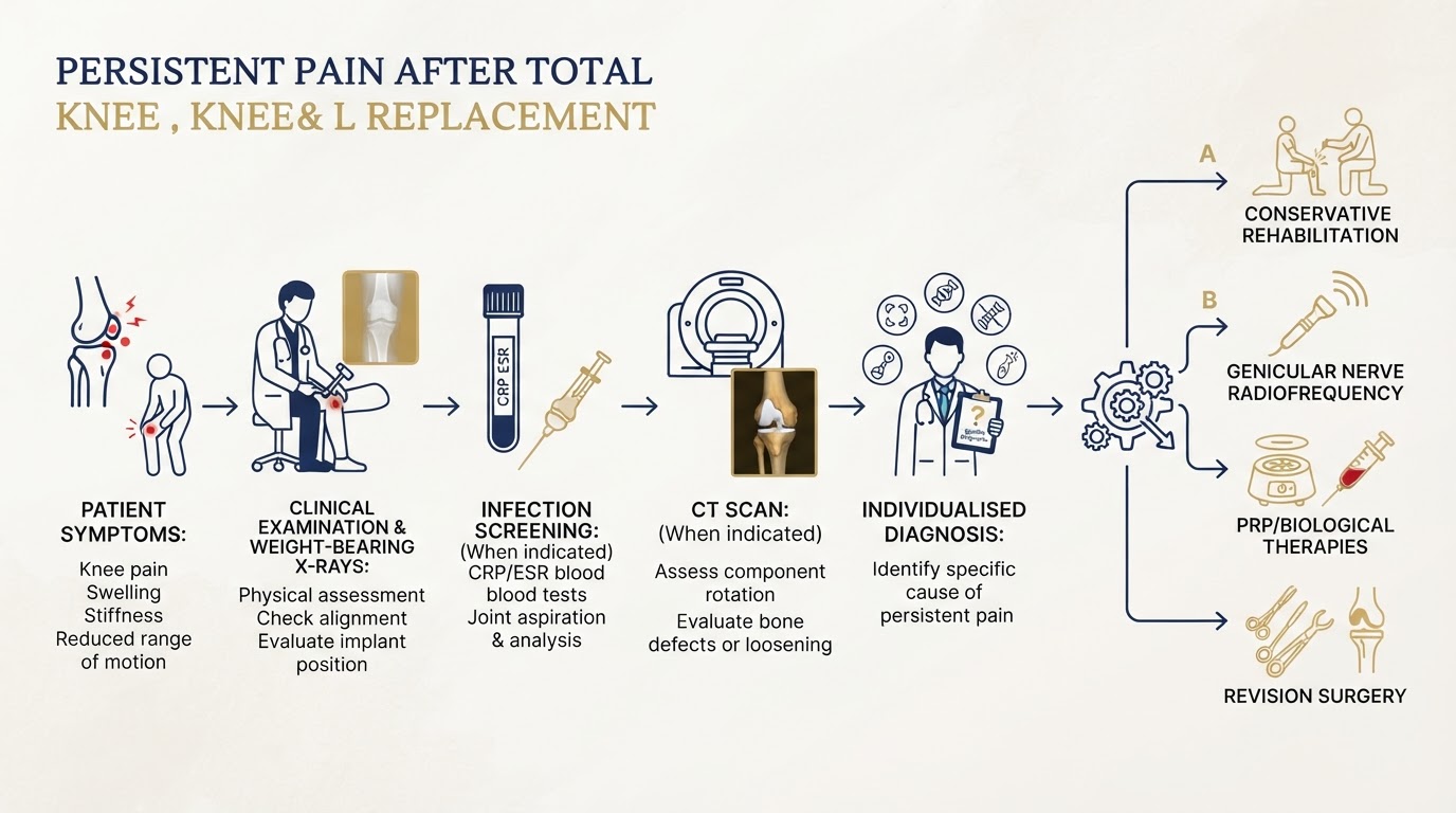
The investigation of persistent pain after knee replacement is systematic and individualised rather than a fixed protocol. The pathway, illustrated in the diagnostic diagram below, typically progresses through several stages: a clinical history and examination (timing, quality and triggers of the pain; change from the postoperative baseline; instability, stiffness or systemic features; palpation of tendons and bursae; hip and spine examination); weight-bearing radiographs with comparison to previous imaging (the single most useful investigation, showing migration, radiolucencies and wear over time); blood tests (CRP and ESR) to screen for infection when indicated; joint aspiration and synovial analysis when screening suggests infection; CT when component rotation, malposition or bone defects need detailed assessment; and selected nuclear medicine studies in equivocal cases. The diagnostic table below connects the main causes with their typical findings, investigations and treatment approach. The aim is to move from symptoms to a working diagnosis and then to an individualised treatment plan — not a generic pathway. Infection and mechanical causes are considered and excluded before pain is attributed to a neuropathic or soft-tissue cause. Diagnostic guide — cause, typical findings, investigations and treatment: * Aseptic loosening — progressive activity-related aching, radiolucencies or migration on weight-bearing films — radiographs with comparison, CT if needed — monitoring or revision when symptomatic and progressive. * Periprosthetic infection — new/night pain, recurrent effusions, wound or systemic features — CRP, ESR, joint aspiration — antibiotics and staged surgical management. * Polyethylene wear / osteolysis — gradual change, instability, lysis on films — radiographs, CT — monitoring or revision depending on severity. * Instability — giving way on stairs or turning, recurrent effusions — examination, radiographs, CT for rotation — rehabilitation or revision/insert exchange. * Patellofemoral pain — anterior pain, catching, maltracking — sunrise view, CT rotation — physiotherapy or selected patellar surgery. * Soft-tissue / tendinopathy — focal tenderness, pain on palpation, normal implant — clinical examination, ultrasound — targeted rehabilitation, load management. * Stiffness / arthrofibrosis — firm limit to range, well-fixed implant — examination, radiographs — intensive physiotherapy, manipulation or lysis of adhesions. * Neuropathic / infrapatellar nerve — burning, shooting, allodynia, nerve territory — clinical pattern, nerve blocks — medication, nerve interventions. * Referred hip / spine pain — hip stiffness, neurogenic symptoms — hip and spine examination, imaging — treat the hip or spine. * Pain with normal radiographs — unremarkable films — extended workup (bloods, aspiration, CT) — identify cause; conservative care if none found.
Conservative treatment and targeted rehabilitation
For many patients, the first and most appropriate response to persistent pain is not surgery but a structured, targeted rehabilitation programme. This is true for soft-tissue pain, stiffness, early anterior knee pain, referred pain being addressed at its source, and for patients in whom no structural cause is found. A well-designed programme addresses quadriceps and hip strength, range of motion, gait retraining, balance and functional activity, and is progressed individually rather than to a generic template. Activity modification, weight management, sensible analgesia and sleep management all play a role. The programme is most effective when it is matched to the identified cause — for example, vastus medialis activation and taping for patellofemoral pain, eccentric loading for tendinopathy, or a dedicated stiffness protocol for arthrofibrosis. Conservative care is not a 'do nothing' option: it is an active, supervised process, and it resolves or substantially improves persistent pain in a meaningful proportion of patients without exposing them to the risks of further surgery.
Genicular nerve blocks and radiofrequency treatment
Genicular nerve radiofrequency is an interventional pain option for selected patients with persistent knee pain. The genicular nerves — the superior and inferior medial and lateral genicular nerves — are the sensory nerves around the knee, and radiofrequency uses heat (conventional thermal), cooled radiofrequency, or pulsed energy to modulate the pain signals they carry. The typical pathway is a diagnostic genicular nerve block first: local anaesthetic is placed on the target nerves under imaging guidance, and a meaningful, documented reduction in pain confirms that those nerves contribute to the pain and helps select patients who may benefit from radiofrequency. Cooled radiofrequency allows larger lesion volumes and is widely used; conventional thermal and pulsed radiofrequency are alternatives with different lesion characteristics. Two important cautions apply. First, most of the published randomised and systematic-review evidence for genicular radiofrequency comes from native knee osteoarthritis, not from patients with a knee prosthesis; that evidence cannot be automatically extrapolated to a replaced knee, where the pain sources and anatomy differ. Second, radiofrequency is a pain-management tool, not a treatment for mechanical failure: it does not correct component loosening, infection, malposition, instability or polyethylene wear. It is considered only when mechanical and infective causes have been assessed and addressed, and when a diagnostic block supports a nerve contribution to the pain. Reported benefit in native-knee studies is variable in degree and duration, and not every patient with persistent pain is a candidate. Complications are generally uncommon but can include temporary numbness, bruising and, rarely, worsening pain or injury to adjacent structures.
Infrapatellar branch of the saphenous nerve interventions
When persistent pain is focal, neuropathic and follows the territory of the infrapatellar branch of the saphenous nerve — typically medial or anterior knee pain with burning or paraesthetic qualities, sometimes related to a scar or a neuroma — targeted interventions on this nerve may be considered. The approach is usually staged: clinical identification of the pain pattern and a diagnostic nerve block to confirm the nerve's contribution; ultrasound-guided injection of local anaesthetic and corticosteroid for short-term relief and diagnostic confirmation; and, in selected cases with a clearly positive block but limited duration of benefit, pulsed or conventional radiofrequency of the infrapatellar branch, or surgical management of a symptomatic neuroma. As with genicular radiofrequency, these are selective options for a specific, identified neuropathic pain source — not a treatment for a loose, infected or malpositioned implant. The evidence base is more limited than for the genicular nerves, and outcomes vary; the decision is made case by case after mechanical and infective causes have been excluded and a diagnostic block supports the nerve as the pain generator.
PRP/PRGF and other biological treatments: current evidence and limitations
Platelet-rich plasma (PRP) and plasma rich in growth factors (PRGF) are biological preparations derived from the patient's own blood, used for their potential anti-inflammatory and regenerative effects. In the context of a knee replacement, three distinct potential applications must be considered separately, because the evidence is very different for each. First, intra-articular PRP in a knee that already contains a prosthesis: there is little published, good-quality clinical evidence specifically examining PRP injected into a replaced knee. Most PRP evidence comes from native knee osteoarthritis, and those results cannot be extrapolated to a prosthetic joint, where the biology, the presence of metal and polyethylene, and the risk of introducing infection are all different. Second, periarticular PRP — injected into tendons, bursae or soft tissues around the replacement — may be considered for an identified soft-tissue cause such as a tendinopathy, on the same principles as in a native knee, but again the specific evidence in replaced knees is limited. Third, there is no reliable clinical evidence that PRP repairs, reattaches or stabilises a loose prosthetic component, or that it reverses aseptic loosening. A critical distinction must be stated plainly: pain relief after a biological injection is not proof of mechanical implant stability, and it does not mean a loose component has become fixed. PRP does not re-fix a loose implant, treat an infection, or correct malposition. Where the evidence for a specific application is insufficient, that should be stated honestly rather than implied. Biological treatments are therefore presented as selective, adjunctive options for specific soft-tissue or inflammatory pain sources — not as a treatment for implant failure — and are used only after infection has been excluded, because injecting a potentially infected joint is harmful.
Other interventional pain-management options
Beyond nerve radiofrequency and biological therapies, several other interventional options form part of a comprehensive pain-management approach when a structural cause has been addressed or excluded. These include ultrasound-guided corticosteroid injection for an identified inflammatory soft-tissue cause (such as bursitis) once infection is excluded; targeted physiotherapy-based desensitisation for neuropathic and persistent postsurgical pain; optimised oral medication, including neuropathic agents where a neuropathic component is identified; and psychological and sleep support, which are recognised components of persistent-pain management. These options are complementary rather than alternative — they are combined according to the individual pain mechanism, and they are most effective when coordinated with the treating surgeon and a pain or rehabilitation specialist. The principle is always the same: interventions are matched to the identified cause, and they do not substitute for treating a mechanical or infective problem when one is present.
When revision surgery is indicated
Revision surgery is not the default response to persistent pain; it is indicated when a defined, surgically correctable cause has been identified and the patient is fit and the expected benefit justifies the risk. Indications include symptomatic aseptic loosening that is progressive, periprosthetic infection, symptomatic instability or malposition that conservative care cannot resolve, significant polyethylene wear with osteolysis or impending failure, and selected patellar or periprosthetic problems. Revision is generally a larger and more complex operation than the primary replacement: it involves removing failed components while preserving bone, reconstructing bone defects, and frequently using stems, augments or cones; recovery is more variable and often slower. The decision is built on the diagnosis, the bone and soft-tissue quality, the patient's general health and expectations, and the surgeon's experience. Crucially, revision is not offered for pain without an identified cause, and it is not offered when the pain is neuropathic, referred, or soft-tissue related and the implant is well-functioning — in those cases, non-surgical management is both safer and more effective.
Conclusion and patient consultation CTA
Persistent pain after a knee replacement is a real and common problem, but it is not a single diagnosis and it does not automatically mean the operation has failed. The right approach is a systematic assessment that distinguishes expected recovery from persistent pain, considers and excludes mechanical and infective causes before attributing pain to a nerve or soft tissue, examines the hip and spine, and builds an individualised treatment plan around the actual cause — whether that is targeted rehabilitation, neuropathic medication, genicular or infrapatellar nerve intervention, a biological therapy for a specific soft-tissue problem, or revision surgery for a defined, correctable implant issue. For residents and visitors across the Costa Blanca — Alicante, Torrevieja and Orihuela Costa — Dr Pablo Banchero offers individualised, evidence-based assessment of persistent pain after knee replacement, whether you are seeking a diagnosis for the first time or a second opinion about a painful prosthesis.
Identify the cause, then match the treatment
Persistent pain after knee replacement has many causes, only some of which involve the implant. The pathway moves from a careful history and examination through targeted imaging and laboratory tests to an individualised diagnosis, and then to a treatment matched to that cause — conservative, interventional or surgical. Radiofrequency and PRP are selective options for specific pain sources, not treatments for a loose or infected implant, and evidence from native knee osteoarthritis is not extrapolated to a replaced knee. On the Costa Blanca — Alicante, Torrevieja and Orihuela Costa — Dr Pablo Banchero provides individualised orthopaedic assessment and second opinions for painful knee replacements.
References & scientific evidence
- American Academy of Orthopaedic Surgeons (AAOS). Diagnosis and Prevention of Periprosthetic Joint Infections of the Hip and Knee — Evidence-Based Clinical Practice Guideline.. AAOS Clinical Practice Guidelines, 2019.Clinical guidelineView source
- American Academy of Orthopaedic Surgeons (AAOS). Surgical Management of Osteoarthritis of the Knee — Evidence-Based Clinical Practice Guideline (SMOAK2).. AAOS Clinical Practice Guidelines, 2022.Clinical guidelineView source
- National Institute for Health and Care Excellence (NICE). Joint replacement (primary): hip, knee and shoulder. NICE guideline NG157.. NICE Clinical Guidelines, 2020.Clinical guidelineView source
- National Institute for Health and Care Excellence (NICE). Chronic pain (primary and secondary) in over 16s: assessment and management. NICE guideline NG193.. NICE Clinical Guidelines, 2021.Clinical guidelineView source
- Parvizi J, Tan TL, Goswami AK, Shea M, Comhey C, Kamath AF, et al.. The 2018 Definition of Periprosthetic Hip and Knee Infection: An Evidence-Based and Validated Criteria.. Journal of Arthroplasty, 2018.Observational studyDOI: 10.1016/j.arth.2018.02.058PMID: 29661564View source
- Zimmerli W, Trampuz A, Ochsner PE. Prosthetic-joint infections.. New England Journal of Medicine, 2004.Systematic reviewDOI: 10.1056/NEJMra040168View source
- Evans JT, Walker RW, Evans JP, Blom AW, Sayers A, Whitehouse MR. How long does a knee replacement last? A systematic review and meta-analysis of case series and national registry reports.. The Lancet, 2019.Meta-analysisDOI: 10.1016/S0140-6736(18)32531-5View source
- Riaz O, Aqil A, Husain A, Chishti I, Hill JP, Nicholl A. Periprosthetic fractures around total knee arthroplasty — a review.. Annals of Medicine and Surgery, 2021.Systematic reviewDOI: 10.1016/j.amsu.2021.102598View source
- National Joint Registry (NJR) for England, Wales, Northern Ireland and the Isle of Man. National Joint Registry 21st Annual Report — Hip, Knee, Ankle, Elbow and Shoulder Arthroplasty: Outcomes and Survivorship.. National Joint Registry, 2024.Observational studyView source
- Australian Orthopaedic Association National Joint Replacement Registry (AOANJRR). Hip, Knee & Shoulder Arthroplasty Annual Report 2024.. AOA NJRR, 2024.Observational studyView source
- Swedish Knee Arthroplasty Register (SKAR). The Swedish Knee Arthroplasty Register — Annual Report.. Swedish Knee Arthroplasty Register, 2023.Observational studyView source
- Campbell KA, Saltzman BM, Mascarenhas R, Khair MM, Verma NN, Cole BJ, Romeo AA. Does Intra-articular Platelet-Rich Plasma Injection Provide Symptomatic Relief in Patients With Knee Osteoarthritis? A Systematic Review of the Literature.. American Journal of Sports Medicine, 2015.Systematic reviewDOI: 10.1177/0363546515580437View source
- Treede RD, Jensen TS, Campbell JN, Cruccu G, Dostrovsky JO, Griffin JW, et al.. Neuropathic pain: redefinition and a grading system for clinical and research purposes.. Neurology, 2008.Observational studyDOI: 10.1212/01.wnl.0000282763.29778.59PMID: 19005277View source
- Davis T, Loudermilk E, DePalma M, Hunter C, Lindley D, Patel N, et al.. Prospective, Multicenter, Randomized, Crossover Clinical Trial Comparing the Safety and Effectiveness of Cooled Radiofrequency Ablation with Corticosteroid Injection in the Management of Knee Pain from Osteoarthritis.. Pain Medicine, 2018.Randomised controlled trialDOI: 10.1093/pm/pnx186View source
Frequently asked questions

Dr. Pablo Banchero Lasalvia
Orthopaedic Surgeon · Traumatology Team
- Author:
- Dr. Pablo Banchero Lasalvia
- Medical reviewer:
- Dr. Pablo Banchero Lasalvia
- Published:
- 2 November 2026
- Medical review date:
- 2 November 2026
- ·
- 22 min read
Book Your Consultation via WhatsApp
Request a private orthopaedic assessment with Dr Pablo Banchero for persistent pain after knee replacement, neuropathic or nerve-related pain, or an expert second opinion before revision surgery on the Costa Blanca.
Official number: +34 602 99 22 22