
Hip replacement is one of the most successful operations in orthopaedic surgery, but implants do not last forever and complications can occur. If a hip replacement becomes painful, the most important step is not to plan a new operation — it is to identify exactly what is wrong. Pain after a hip replacement does not automatically mean the implant has failed, and revision surgery is not always the right answer. This article explains why hip replacements can fail, how the cause is diagnosed, when revision surgery is genuinely necessary, when it can be avoided, and what reconstructive options exist. At TraumatologyTeam, on the Costa Blanca — Alicante, Torrevieja, Orihuela Costa and La Zenia — we approach revision as a decision built on a precise diagnosis and an individualised reconstructive plan, not as a routine exchange of components.
What Is Revision Hip Replacement Surgery?
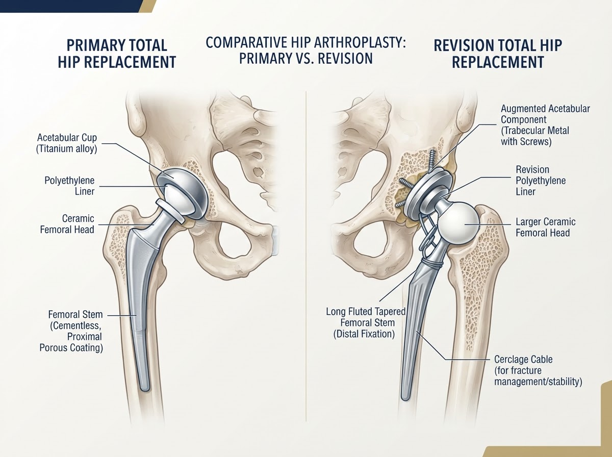
Revision hip replacement is an operation that replaces, modifies or reconstructs one or more components of a previous hip replacement. It differs from a primary total hip replacement in both purpose and complexity. • Primary total hip replacement: the first operation to replace the diseased hip joint with artificial components. • Revision total hip replacement: a re-operation on a hip that already has a prosthesis, performed to correct a problem with the implant, the bone, the soft tissues or an infection. • Component exchange: replacing one part of the implant while keeping others. • Partial revision: replacing only the femoral component or only the acetabular component. • Complete revision: replacing both the femoral and acetabular components. Revision surgery may involve removing well-fixed but problematic components, reconstructing bone that has been lost, dealing with infection, or correcting instability. Because each revision is different, the procedure must be planned around the specific reason the previous reconstruction has failed. PRIMARY VS REVISION HIP REPLACEMENT: • Bone: Primary = native bone available; Revision = bone loss common, may need reconstruction. • Components: Primary = standard components; Revision = often long stems, augments, custom or cup-cage constructs. • Surgical time: Primary = usually shorter; Revision = typically longer and more complex. • Risk: Primary = lower; Revision = higher infection, dislocation, fracture and re-revision risk. • Recovery: Primary = generally faster; Revision = often slower and more individualised.
Does Every Painful Hip Replacement Need Revision?
No — and this is one of the most important messages in this article. Pain after a hip replacement can come from many sources, and only some of them require revision surgery. Pain may be related to the implant (loosening, wear, malposition or infection), but it may also come from structures around the joint: • Iliopsoas tendinopathy (irritation of the psoas tendon as it passes in front of the acetabular cup). • Trochanteric pain or bursitis on the outside of the hip. • Referred pain from the lumbar spine. • Muscle weakness or imbalance. • Extra-articular causes such as sacroiliac joint pain or nerve compression. • Occult periprosthetic fracture. Pain after hip replacement does not automatically mean implant failure. Investigating the cause before recommending a new operation protects patients from unnecessary revision surgery. When the source of pain is extra-articular, the correct treatment may be physiotherapy, medication, a targeted injection or management of the spine — not another hip operation.
Why Do Hip Replacements Fail?
Hip replacements can fail for several reasons, and identifying the mechanism of failure is the foundation of any revision plan. The principal causes are: • Aseptic loosening — loss of fixation without infection, often driven by wear particles and osteolysis. • Periprosthetic joint infection — infection involving the implant, which may be acute or chronic. • Recurrent dislocation — repeated instability of the prosthesis. • Polyethylene wear and osteolysis — progressive loss of bearing material and bone resorption. • Periprosthetic fracture — a break in the bone around the implant. • Implant breakage — mechanical failure of a component. • Adverse local tissue reactions — inflammatory response to metal debris or modular corrosion. • Persistent instability — recurrent dislocation despite intact components. • Component malposition — poor orientation causing impingement, wear or instability. • Other causes of persistent pain without mechanical failure. CAUSES OF HIP REPLACEMENT FAILURE: • Aseptic loosening: Loss of fixation without infection. Often wear-mediated. Diagnosed with radiographs, confirmed at surgery. • Infection: Biofilm on the implant. May present with or without fever. Requires combined medical and surgical strategy. • Dislocation: Femoral head leaves the cup. May recur if position, soft-tissue balance or anatomy is unfavourable. • Wear and osteolysis: Polyethylene wear particles provoke bone resorption around the implant, often silently. • Periprosthetic fracture: Break around the implant after a fall or from fragile bone. Classification guides treatment. • Component malposition: Poor cup or stem position causes impingement, instability or accelerated wear. • Adverse local tissue reactions: Inflammatory reaction to debris, more common with some historical metal-on-metal designs. • Implant breakage: Rare with modern materials, but possible in stems or liners under high load.
Aseptic Loosening
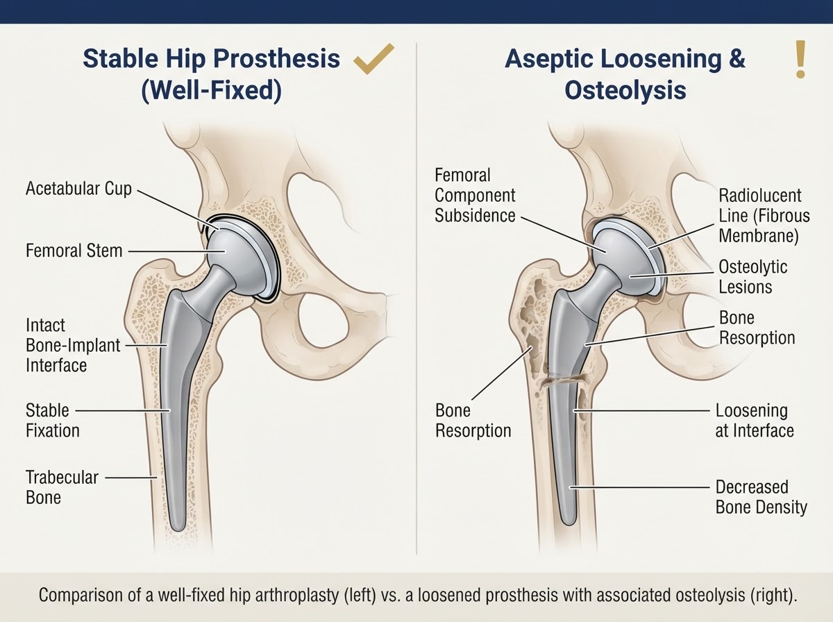
Aseptic loosening means the implant has lost its fixation to bone in the absence of infection. It is one of the most common late reasons for revision. It may affect the femoral stem, the acetabular cup, or both. The process is often driven by polyethylene wear particles that provoke osteolysis — progressive resorption of bone around the implant. As bone is lost, the component loses support, may migrate, and becomes painful. Radiographs may show radiolucent lines around the implant, migration, or lytic areas where bone has been resorbed. Not every radiographic change means failure. Some stable changes are expected and do not require intervention. The distinction that matters is between stable radiographic findings and progression that suggests the component is loosening. When loosening becomes symptomatic, with pain on weight-bearing or progressive loss of bone, revision is usually indicated before the bone loss becomes severe enough to make reconstruction far more difficult.
Periprosthetic Joint Infection
Periprosthetic joint infection (PJI) is infection involving the implant. It is one of the most serious complications because bacteria form a biofilm on the implant surface that is difficult to clear with antibiotics alone. Infections are classified by how and when they appear: • Early infection: appearing within weeks of surgery, often acute and with obvious signs. • Delayed or chronic infection: appearing months later, sometimes with subtle symptoms. • Haematogenous late infection: caused by bacteria reaching the implant through the bloodstream from a distant source, sometimes years after surgery. An important point for patients: periprosthetic infection can exist without a high fever or obvious wound redness. Low-grade, chronic infection may cause persistent aching pain, night pain, or repeated episodes of inflammation that settle with antibiotics and return. This is why infection must be actively investigated before any revision surgery, even when the clinical picture seems mild. Treating a loose, infected hip as if it were aseptic loosening is one of the most serious errors in revision surgery.
Recurrent Hip Dislocation and Instability
Dislocation occurs when the femoral head comes out of the acetabular cup. When it happens repeatedly, it becomes a disabling problem rather than a one-off event. Recurrent dislocation may be caused by: • Component malposition — cup inclination or anteversion, or femoral version, outside a stable range. • Impingement — bony or component contact that levers the head out of the cup. • Muscle deficiency or abductor insufficiency. • Neuromuscular conditions affecting muscle control. • Soft-tissue imbalance, including reduced offset. • Spinopelvic abnormalities affecting the functional position of the pelvis. The management of instability depends on identifying its cause. Correcting component position, restoring offset, repairing soft tissues where possible, and selecting larger heads, dual mobility or, in complex cases, constrained liners are all options — each with its own advantages and limitations. This topic connects directly with Article 18 (surgical approaches), Article 19 (implant selection and bearing surfaces) and Article 20 (complications and prevention).
Polyethylene Wear and Osteolysis
Polyethylene is the most common liner material. Over time it wears, generating small particles. The body's inflammatory response to these particles can drive osteolysis — progressive resorption of bone around the implant. A characteristic feature of this process is that significant osteolysis can develop before the patient feels severe pain. The bearing may be wearing and the bone may be quietly disappearing while the hip still feels reasonably comfortable. This is one reason long-term radiographic follow-up matters: detecting wear and osteolysis early allows revision to be planned while there is still enough bone to reconstruct reliably. Modern highly cross-linked polyethylene and ceramic bearings have substantially reduced wear compared with earlier materials, but no bearing is immune to time and load. When wear and osteolysis become significant, revision of the affected components is usually indicated to prevent further bone loss and eventual failure.
Periprosthetic Femoral and Acetabular Fractures
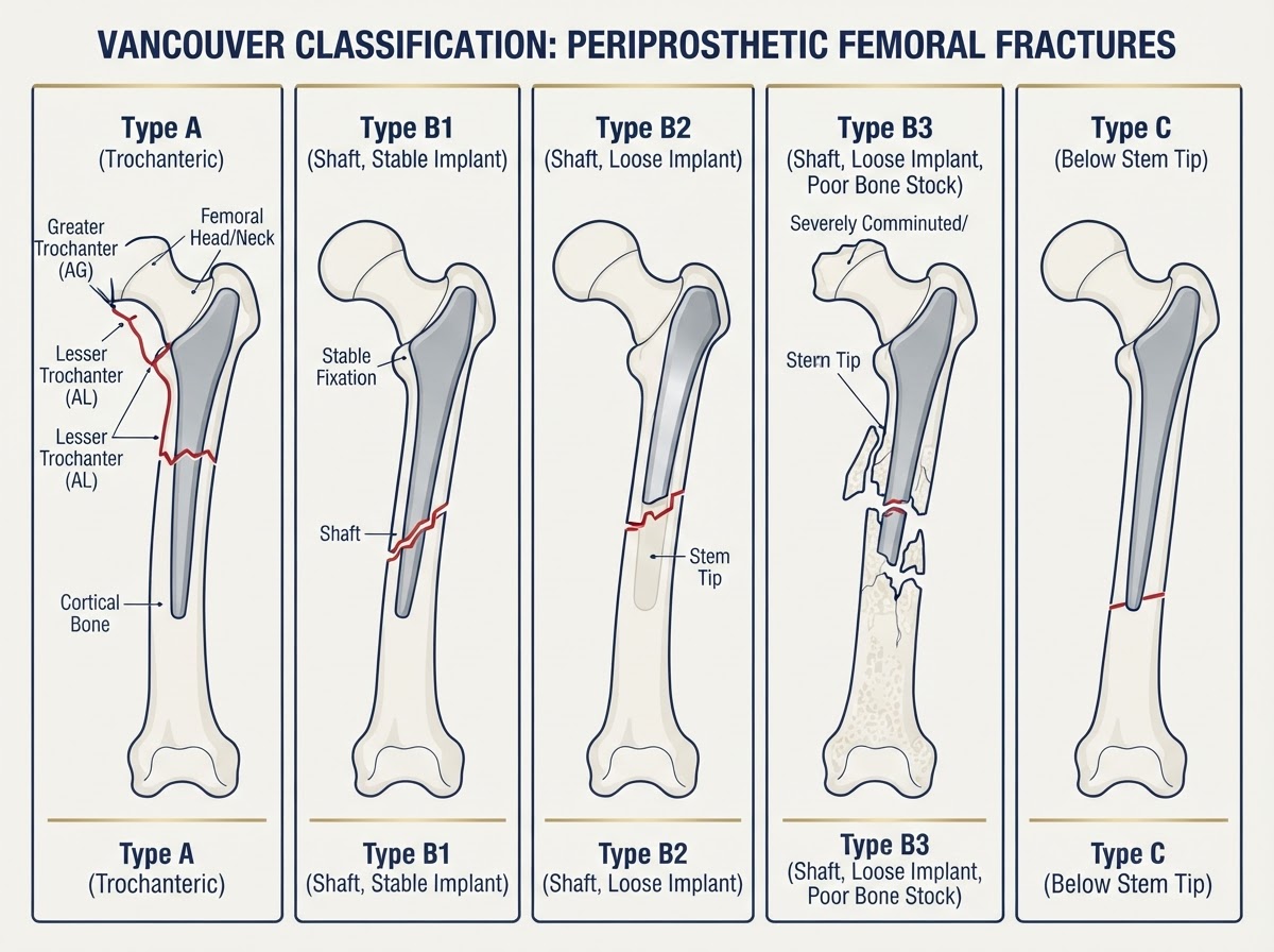
A periprosthetic fracture is a break in the bone around an implant. It can occur during surgery (intraoperative) or afterwards (postoperative), and it may affect the femur or, less commonly, the acetabulum. For femoral periprosthetic fractures, the Vancouver classification is widely used because it links the fracture pattern to implant stability and so guides treatment: • Type A: fractures around the trochanteric region (greater or lesser trochanter). • Type B1: fractures around the stem with a well-fixed, stable stem. • Type B2: fractures around the stem with a loose stem. • Type B3: fractures around the stem with a loose stem and poor bone stock. • Type C: fractures below the tip of the stem, with the stem itself well-fixed. The key distinction is whether the stem is still firmly fixed. A fracture around a stable stem (B1) may be treated with fixation alone, while a fracture around a loose stem (B2 or B3) requires revision of the stem — usually with a long stem that bypasses the fracture and achieves fixation beyond it. This single distinction changes the entire operation and outcome, which is why fracture classification matters.
Adverse Local Tissue Reactions and Trunnionosis
Adverse local tissue reactions are inflammatory responses that can occur around a hip replacement, most often linked to corrosion at the modular junction between the femoral head and stem (trunnionosis) or to historical metal-on-metal bearing surfaces. Metal debris from corrosion or wear can provoke a soft-tissue reaction that damages muscle, tendon and bone. With some older metal-on-metal designs, this led to pseudotumour formation and significant tissue injury. Evaluation depends on the type of implant, the patient's symptoms, blood metal-ion levels when indicated, and cross-sectional imaging (MRI with metal-artefact reduction) to assess the soft tissues around the joint. When an adverse local tissue reaction is confirmed and is causing symptoms or progressive tissue damage, revision of the offending bearing or component is usually indicated. Not every elevated metal ion or radiographic reaction requires revision; the decision integrates symptoms, imaging and the natural history of the specific implant.
How Is a Failed Hip Replacement Diagnosed?
Diagnosing a failing hip replacement is a structured process. No single test rules out every cause; the diagnosis is built from the combination of findings. CLINICAL ASSESSMENT: • Pain and its location (groin, thigh, trochanter, buttock). • Onset and evolution — did it start suddenly or progress slowly? • Pain on weight-bearing versus pain at rest or at night. • Fever or inflammatory signs. • History of dislocation episodes. • Function and mobility, including any decline. • Previous operations and implant details. IMAGING: • Anteroposterior pelvic radiograph and specific hip projections. • Comparison with previous films to detect migration, radiolucent lines or osteolysis. • CT for bone loss, component position and surgical planning. • MRI with metal-artefact reduction when soft-tissue reactions are suspected. • Other studies as indicated by the clinical question. LABORATORY ASSESSMENT: • Blood count, C-reactive protein (CRP) and erythrocyte sedimentation rate (ESR). • Joint aspiration with cell count and cultures when infection is suspected. • Synovial fluid analysis. No single test excludes every cause of failure. The diagnosis is a clinical synthesis, and in difficult cases it may require several steps before the mechanism is understood.
Diagnosing Infection Before Revision Surgery
Because infection changes the entire surgical strategy, ruling infection in or out before revision is essential. The workup commonly combines: • ESR and CRP — inflammatory markers that, when normal, help exclude infection and, when elevated, raise suspicion. • Synovial white blood cell count — the number of inflammatory cells in the joint fluid. • Synovial neutrophil percentage — the proportion of a specific white cell type. • Cultures — to identify the responsible organism and guide antibiotic therapy. • Alpha-defensin in selected situations — a synovial marker that supports the diagnosis of infection. • Intraoperative histology, when appropriate, to confirm acute inflammation. The International Consensus Meeting (ICM) and Musculoskeletal Infection Society (MSIS) criteria combine these findings into a structured definition of periprosthetic joint infection, and they are widely used to guide decisions. These criteria are not infallible: low-grade infection can be difficult to diagnose and may be missed even with a full workup. There is an important difference between a confirmed diagnosis of infection and clinical suspicion, and in uncertain cases the decision may be made with the help of a second opinion from a revision specialist.
When Is Revision Surgery Necessary?
Revision is usually indicated when there is a clear, identifiable problem that conservative treatment cannot resolve: • Symptomatic and progressive aseptic loosening. • Confirmed or high-suspicion periprosthetic joint infection. • Recurrent dislocation that is not controlled by non-operative measures. • Advanced wear with significant osteolysis, even when still relatively painless, to preserve bone. • Periprosthetic fracture around a loose or unstable implant. • Mechanical failure of a component. • Significant soft-tissue damage related to the implant. The decision integrates symptoms, functional impairment, radiographic findings and the risks of the operation. A patient with a clearly failing hip and meaningful loss of function should not be made to exhaust every possible injection before surgery is offered. Equally, revision should not be recommended when the diagnosis is uncertain or the symptoms can be managed another way.
When Can Revision Be Avoided?
In some situations, revision is not the right answer even though the hip hurts. Revision can be avoided, or at least postponed, when the pain has a treatable cause that does not involve the implant: • A well-fixed, well-positioned implant with no signs of progression. • Pain of lumbar spinal origin. • Iliopsoas tendinopathy, which may respond to activity modification, physiotherapy or a targeted injection. • Trochanteric pain or bursitis. • Muscle-related pain or weakness responsive to rehabilitation. • Functional or biomechanical problems that can be addressed without surgery. • Incidental radiographic findings without clinical correlation. Correctly identifying the cause avoids unnecessary surgery. The value of a careful diagnostic process is precisely this: it distinguishes a hip that genuinely needs revision from one that needs a completely different treatment.
Partial Revision vs Complete Revision
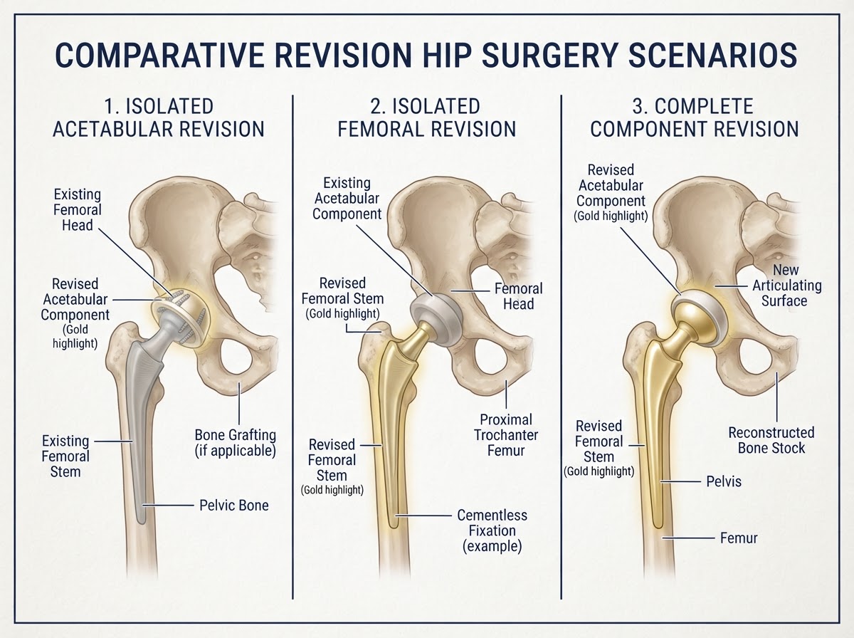
When revision is indicated, the scope of the operation must match the mechanism of failure. Not every revision requires replacing everything. PARTIAL REVISION: • Isolated femoral component revision — when the acetabular side is well-fixed, well-positioned and healthy. • Isolated acetabular component revision — when the femoral stem is solid and the cup is the problem. • Liner exchange — replacing a worn liner while keeping the metal shell, when the shell is stable and compatible. • Head and liner exchange — exchanging the bearing surfaces while retaining well-fixed components. COMPLETE REVISION: • Replacement of both the femoral and acetabular components, used when both sides are loose, infected, or when the problem involves the whole reconstruction. A component can be retained only when it is well-fixed, correctly positioned, structurally intact, compatible with the new components, and there is no infection forcing a different strategy. Partial revision is not automatically preferable: keeping a component that is subtly loose or malpositioned risks early re-revision. The choice depends entirely on what is found, not on a preference for a shorter operation. PARTIAL VS COMPLETE REVISION: • Indication: Partial = one side is healthy and stable; Complete = both sides failed or one side dictates full revision. • Bone preservation: Partial = preserves fixed bone and components; Complete = may require reconstruction of both sides. • Complexity: Partial = generally lower; Complete = higher, longer surgery. • Risk: Partial = usually lower; Complete = higher infection, dislocation and re-revision risk. • Decision driver: Partial = stable opposite side; Complete = infection, both-side loosening, or severe bone loss.
Revision Femoral Stems
When the femoral component must be revised, the stem chosen has to achieve stable fixation in the remaining bone. Options include: • Long stems — bypass areas of bone loss or fracture and achieve fixation further down the femur. • Distal fixation stems — rely on intact diaphyseal bone when the proximal metaphysis is deficient. • Modular revision stems — allow adjustment of length, offset and anteversion in complex anatomy. • Tapered fluted stems — engage the inner femoral cortex over a long segment. • Cemented revision stems — used in selected cases, including when bone quality or anatomy favours cement. • Other reconstructive systems for specific patterns of bone loss. The principle is to obtain reliable fixation where healthy bone remains. When proximal bone is lost, fixation moves distally; when the canal is wide and thin, the design and technique adapt accordingly. No single stem is universally suitable. Selection depends on the bone available, the reason for revision, the patient's anatomy and the surgeon's experience.
Acetabular Reconstruction
Acetabular revision is needed when the cup is loose, infected, malpositioned or associated with bone loss. Reconstructive options include: • Revision acetabular shells — components designed to gain fixation in compromised bone. • Highly porous metal components — encouraging bone ingrowth where cancellous bone is poor. • Augments — metal pieces that fill segmental defects so the cup can be supported. • Structural bone grafts — replacing lost bone with graft to restore the socket. • Trabecular metal — a porous tantalum material that supports bone ingrowth. • Custom triflange components — designed from CT to fit severe, complex defects. • Cup-cage constructs — combining a cage with a cup for large defects. The Paprosky classification grades acetabular bone loss according to the remaining support for a new cup, helping the surgeon plan the level of reconstruction needed. The aim is to restore a stable, well-positioned socket that can support the new bearing — without overcomplicating the procedure when a simpler reconstruction would be sufficient.
Managing Severe Bone Loss
Severe bone loss is the central challenge in complex revision. It may affect the femur, the acetabulum, or both, and it takes different forms: • Segmental defects — loss of a wall or section of bone. • Cavitary defects — loss of the inner bone filling. • Structural instability — the remaining bone can no longer support an implant in the original position. Reconstructive planning must consider how much healthy bone remains and where stable fixation can be obtained. The goal is to restore biomechanics with the least destructive strategy that will be durable — sometimes a bone graft, sometimes an augment, sometimes a custom or cage construct, and sometimes a combination. Severe bone loss makes revision longer, riskier and less predictable, which is why early detection of loosening and wear, before bone is lost, is so important.
Revision for Recurrent Dislocation
When dislocation recurs despite non-operative management, revision may be needed to correct the underlying cause. Options include: • Correction of component malposition — repositioning a cup or stem that is causing impingement or instability. • Restoring offset — rebuilding soft-tissue tension around the joint. • Soft-tissue repair, when appropriate, to reconstruct the abductor mechanism. • Larger femoral heads — increasing the jump distance in compatible constructs. • Dual mobility — a mobile bearing that increases the range before dislocation can occur. • Constrained liners — used in selected complex or recurrent cases where other measures have failed. Each strategy has advantages and limitations. Dual mobility and constrained liners are not universal solutions: dual mobility can be an excellent choice in selected high-risk patients, while constrained liners trade some range of motion and increased load for stability, and are reserved for situations where standard options have failed or are unsuitable. The right choice depends on the cause of the instability, the state of the components and the patient's individual risk.
Revision Surgery for Infection: One-Stage and Two-Stage Revision
When infection is confirmed or strongly suspected, the strategy changes. The main approaches are: DAIR (Debridement, Antibiotics and Implant Retention): • Used in selected early or acute infections where the implant is well-fixed and the tissues are healthy. • Components are retained; modular parts (head and liner) may be exchanged. • Combined with targeted antibiotic therapy. ONE-STAGE REVISION: • Removal and replacement of the infected components in a single operation. • Reserved for carefully selected patients with known, susceptible organisms and adequate soft tissues. TWO-STAGE REVISION: • First stage: removal of the implant, thorough debridement and placement of an antibiotic spacer in selected cases. • A period of antimicrobial therapy. • Second stage: reimplantation once infection is considered controlled. The choice depends on the organism, the state of the tissues, the patient's overall condition and the experience of the surgical team. No single strategy is superior for every patient. In complex or resistant infection, two-stage revision is often chosen, but one-stage revision is a valid option in well-selected cases with experienced teams. ONE-STAGE VS TWO-STAGE REVISION: • Operations: One-stage = single procedure; Two-stage = two procedures with an interval. • Patient selection: One-stage = defined organism, good tissues; Two-stage = complex, resistant or recurrent infection. • Antibiotic interval: One-stage = perioperative; Two-stage = extended course between stages. • Recovery: One-stage = single recovery; Two-stage = longer overall, with a period of limited mobility. • Reimplantation: One-stage = immediate; Two-stage = when infection is considered controlled.
What Are the Risks of Revision Hip Surgery?
Revision hip replacement is generally more complex than a primary hip replacement, and the risks are higher. Potential complications include: • Infection — including recurrence or new infection. • Dislocation — more common in revision because of altered anatomy and soft-tissue tension. • Periprosthetic fracture — during surgery or afterwards. • Greater blood loss — because the operation is longer and exposes more bone. • Neurovascular injury — to the sciatic, femoral or other nerves. • Venous thromboembolism — deep vein thrombosis or pulmonary embolism. • Recurrent loosening — the revised implant can loosen again over time. • Wound-healing problems. • The need for a further revision. Generic risk percentages are unhelpful without specifying the population, the indication and the length of follow-up. The risk for an isolated liner exchange is not the same as for a complete revision with bone reconstruction. A realistic discussion of risk must be tailored to the individual patient and the planned procedure.
Recovery After Revision Hip Replacement
Recovery after revision is influenced by why the operation was done and how the hip was reconstructed. In general terms, recovery commonly involves: • A hospital stay, the length of which depends on the complexity of the reconstruction. • Pain control adapted to the procedure. • Early mobilisation, where the reconstruction allows. • Physiotherapy tailored to the operation and the patient's condition. • Weight-bearing and activity guided by the type of reconstruction — some revisions need protected weight-bearing for a period. • Walking aids as required. • Thromboprophylaxis according to individual risk. • Radiographic follow-up to confirm stable fixation and healing. Recovery may be slower than after a primary hip replacement, and it does not follow a single universal timetable. Returning to activity is guided by the surgeon according to the specific reconstruction and how healing progresses.
Can a Revision Hip Replacement Last as Long as a Primary Hip Replacement?
The survival of a revision hip replacement depends on several factors, and it is generally lower than that of a well-performed primary replacement. Outcomes depend on: • The reason for revision — infection and instability carry different risks than simple wear. • Bone quality and the amount of bone loss at the time of revision. • The number of previous revisions. • The type of reconstruction used. • The patient's age and functional status. • Whether infection was present. • The quality of the fixation achieved. National joint registries show that revision surgery can be durable, but the re-revision rate is higher than the revision rate after primary replacement. No honest surgeon can offer a guaranteed lifespan for a revised hip. What can be said is that a well-indicated, well-planned and well-executed revision, with appropriate reconstruction and follow-up, can provide many years of reliable function — and that the earlier a problem is identified, before severe bone loss, the better the prospects for a durable reconstruction.
The Importance of a Specialist Second Opinion
A second opinion can be valuable at several points in the care of a painful hip replacement: • Persistent pain without a clear diagnosis. • Suspicion of loosening on radiographs. • A recommendation for complete revision when it is unclear whether one component could be retained. • Repeated dislocations. • Suspicion of infection. • Significant bone loss that may require complex reconstruction. • A recommendation for revision when the patient is uncertain whether surgery is truly necessary. The central message: before undergoing revision surgery, it is essential to understand exactly what is failing and which components actually need to be replaced. A specialist assessment offers the opportunity to review the diagnosis, the imaging, the surgical history and the available alternatives. This is not about discrediting another surgeon's recommendation; it is about making sure the decision is based on a precise diagnosis before a second operation is undertaken.
Our Approach to Complex Hip Problems
At TraumatologyTeam, on the Costa Blanca — Alicante, Torrevieja, Orihuela Costa and La Zenia — we approach complex hip problems with a structured process: • Individualised clinical assessment of the painful hip replacement. • Review of imaging and previous surgical records, including the implant in place. • Identification of the mechanism of failure — mechanical, infectious, biological or extra-articular. • Assessment of fixation and bone loss. • Investigation of infection when indicated. • Reconstructive planning matched to the individual anatomy and bone stock. • Transparent discussion of risks, alternatives and expected outcomes. Our philosophy is that revision should be performed only when there is a clear clinical reason, and that the scope of the operation should match the problem — no more and no less. We do not claim to perform every type of complex reconstruction, and when a case requires a highly specialised revision centre, we say so and refer appropriately. The distinction between a specialist assessment and the treatments we actually offer is explained honestly to each patient.
Final Clinical Perspective
Revision hip replacement surgery is not simply about replacing an old implant. It is about identifying why the previous reconstruction has failed and developing a strategy to restore stability, function and comfort while preserving as much healthy bone as possible. The most important decision is not only which prosthesis to implant, but correctly identifying the cause of the problem and choosing the intervention that fits that cause. A painful hip replacement does not automatically need revision; when it does, the right operation may be a partial or a complete revision, an infection strategy, or a reconstruction designed around the bone that remains. The best results come from a precise diagnosis, an individualised reconstructive plan, and honest, shared decision-making between surgeon and patient.
The right revision begins with the right diagnosis
At TraumatologyTeam, on the Costa Blanca — Alicante, Torrevieja, Orihuela Costa and La Zenia — we treat a painful hip replacement as a problem to be diagnosed before it is a problem to be operated on. Revision is never routine, and it is never the first answer to pain. When it is genuinely necessary, we plan the reconstruction around the individual cause, the bone that remains and the patient's goals. If you have a painful hip replacement, or you have been told you may need revision surgery, a specialist assessment with Dr Pablo Banchero offers a structured second opinion before any further operation.
References & scientific evidence
- NICE Guideline NG157. Joint replacement (primary): hip, knee and shoulder. Clinical guideline.. National Institute for Health and Care Excellence, 2020.Clinical guidelineView source
- NICE Guideline NG226. Osteoarthritis in over 16s: diagnosis and management.. National Institute for Health and Care Excellence, 2022.Clinical guidelineView source
- AAOS Clinical Practice Guideline. Diagnosis and Prevention of Periprosthetic Joint Infections of the Hip and Knee.. American Academy of Orthopaedic Surgeons, 2019.Clinical guidelineView source
- AAOS/AAHKS Clinical Practice Guideline. Management of Patients Undergoing Elective Hip or Knee Arthroplasty: Dental Interventions and the Prevention of Periprosthetic Joint Infection.. American Academy of Orthopaedic Surgeons, 2024.Clinical guidelineView source
- AAOS Clinical Practice Guideline. Management of Osteoarthritis of the Hip: Evidence-Based Clinical Practice Guideline.. American Academy of Orthopaedic Surgeons, 2017.Clinical guidelineView source
- National Joint Registry (NJR) for England, Wales, Northern Ireland and the Isle of Man. National Joint Registry 21st Annual Report — Hip, Knee, Ankle, Elbow and Shoulder Arthroplasty: Outcomes and Survivorship, including Revision Burden.. National Joint Registry, 2024.Observational studyView source
- Australian Orthopaedic Association National Joint Replacement Registry (AOANJRR). Hip, Knee & Shoulder Arthroplasty: Annual Report 2024 — including Revision Hip Arthroplasty Outcomes.. AOA NJRR, 2024.Observational studyView source
- Swedish Hip Arthroplasty Register (SHPR). Swedish Hip Arthroplasty Register Annual Report — outcomes, reoperations and revisions.. Swedish Hip Arthroplasty Register, 2023.Observational studyView source
- American Joint Replacement Registry (AJRR). American Joint Replacement Registry Annual Report — hip and knee arthroplasty procedures and revisions.. American Joint Replacement Registry, 2023.Observational studyView source
- Parvizi J, Tan TL, Goswami K, et al.. The 2018 Definition of Periprosthetic Hip and Knee Infection: An Evidence-Based and Validated Criteria.. The Journal of Arthroplasty, 2018.Observational studyDOI: 10.1016/j.arth.2018.02.078View source
- Zimmerli W, Trampuz A, Ochsner PE.. Prosthetic-Joint Infections.. New England Journal of Medicine, 2004.Observational studyPMID: 15483283View source
- Duncan CP, Maspi BA.. Fractures of the femur after hip replacement — the Vancouver classification of periprosthetic femoral fractures.. Instructional Course Lectures, 1995.Observational study
- Paprosky WG, Perona PG, Lawrence JM.. Acetabular defect classification and surgical reconstruction: revision of the deficient acetabulum.. Orthopedic Clinics of North America, 1993.Observational study
- Bozic KJ, Kurtz SM, Lau E, et al.. The epidemiology of revision total hip arthroplasty in the United States.. The Journal of Bone and Joint Surgery (American), 2009.Observational studyDOI: 10.2106/JBJS.H.00155View source
- Kurtz SM, Lau E, Watson H, Schmier JK, Parvizi J.. Economic burden of periprosthetic joint infection in the United States.. Journal of Arthroplasty, 2012.Observational studyDOI: 10.1016/j.arth.2012.02.022View source
- Garcia DC, Darrith B, Davies CM, Bohl DD, Bhandari M, Della Valle CJ.. Periprosthetic Joint Infection Following Hip and Knee Arthroplasty: A Review of Diagnosis and Management.. JAMA Surgery, 2024.Systematic reviewDOI: 10.1001/jamasurg.2024.0153View source
- Schwarzkopf R, Baghoolizadeh M, Long WJ, Della Valle CJ.. Dislocation Following Total Hip Arthroplasty: A Comprehensive Review.. Journal of the American Academy of Orthopaedic Surgeons, 2024.Systematic reviewDOI: 10.5435/JAAOS-D-23-00570View source
- Abdel MP, Watts CD, Wuthrich PS, Berry DJ, Hanssen AD.. Outcomes and complications of revision total hip arthroplasty in the setting of periprosthetic femoral fracture.. The Bone & Joint Journal, 2017.Observational studyDOI: 10.1302/0301-620X.99B6.BJJ-2016-0776.R1View source
- Lewinnek GE, Lewis JL, Tarr R, Compere CL, Zimmerman JR.. Dislocations after total hip-replacement arthroplasties.. Journal of Bone and Joint Surgery (American), 1978.Observational studyPMID: 670295View source
- Cichocki AR, Law TY, Rosas S, et al.. Periprosthetic femur fractures after total hip arthroplasty: a systematic review of risk factors.. Orthopedic Reviews, 2019.Systematic reviewDOI: 10.4081/or.2019.7772View source
- Vigdorchik JM, Sharma AK, Elbuluk AM, et al.. Spinopelvic dysplasia and hypermobility increase dislocation risk in total hip arthroplasty: a functional planning protocol.. The Bone & Joint Journal, 2021.Observational studyDOI: 10.1302/0301-620X.103B7.BJJ-2020-2391.R1View source
Frequently asked questions

Dr. Pablo Banchero Lasalvia
Orthopaedic Surgeon · Traumatology Team
- Author:
- Dr. Pablo Banchero Lasalvia
- Medical reviewer:
- Dr. Pablo Banchero Lasalvia
- Published:
- 2 November 2026
- Medical review date:
- 2 November 2026
- ·
- 26 min read
Request a Specialist Assessment via WhatsApp
Concerned about a painful or failing hip replacement? Request a private orthopaedic second opinion with Dr Pablo Banchero to review your diagnosis, imaging and revision options.
Official number: +34 602 99 22 22