
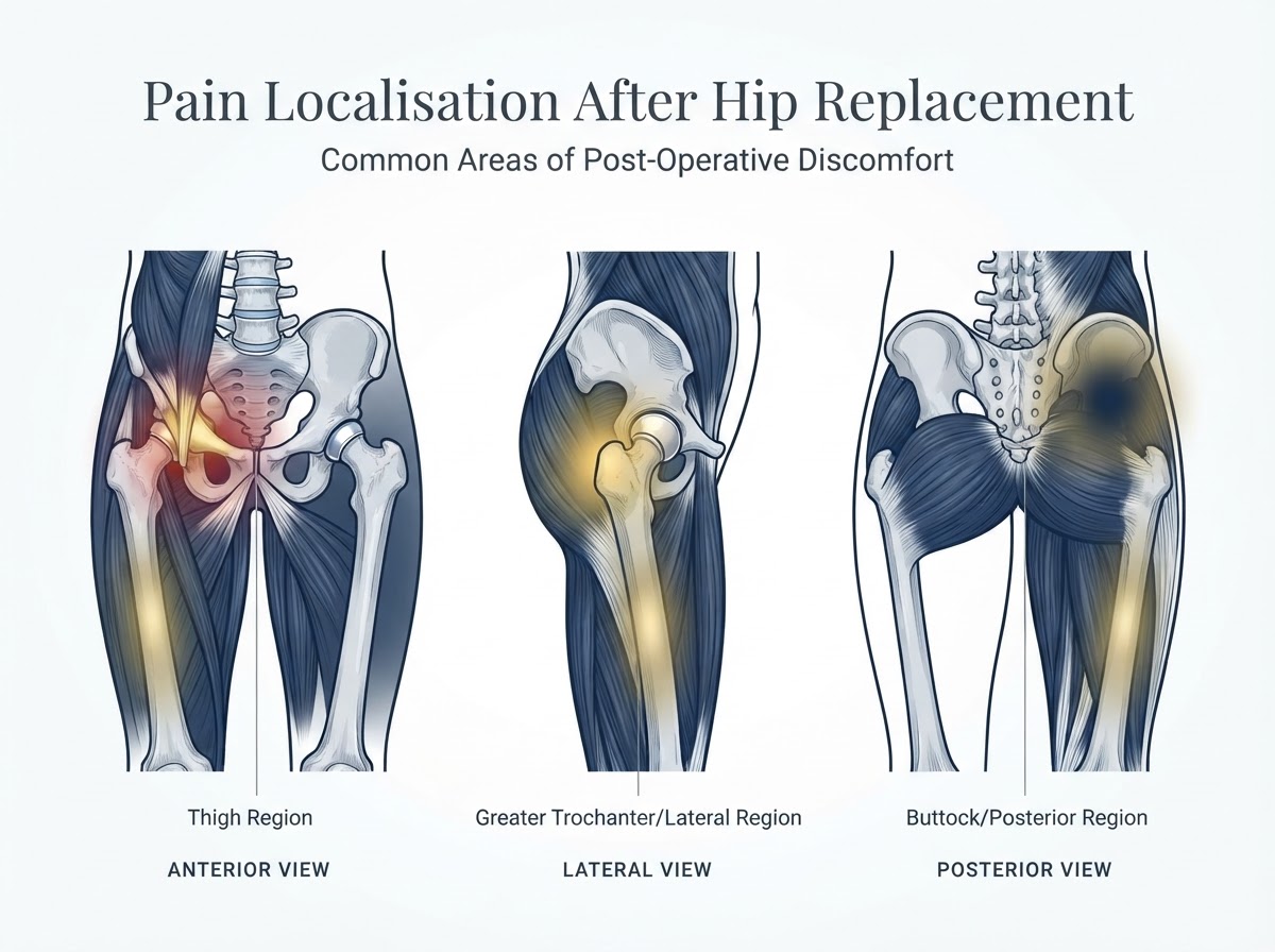
Total hip replacement (total hip arthroplasty) is one of the most effective operations in orthopaedic surgery for relieving the pain of advanced hip osteoarthritis and restoring function. Most patients do well. Yet a meaningful number experience persistent, recurrent or newly developed pain after surgery, and for those patients the experience can be unsettling — particularly the fear that the implant has failed. The central message of this guide is reassuring but precise: pain after hip replacement deserves proper investigation, but it does not automatically mean the implant has failed. The first clinical task is to work out where the pain is coming from — the prosthetic joint itself, the soft tissues around it, the lumbar spine, or another source — because the correct treatment depends entirely on the correct cause. This article walks through how an experienced orthopaedic surgeon reasons through persistent hip pain: how the location and timing of pain guide the differential diagnosis, which investigations are useful and which are not, when infection must be excluded before anything else, and when revision surgery is — and is not — the right answer. It is written for patients considering a specialist assessment or an independent second opinion, and it deliberately avoids attributing symptoms to implant loosening before the cause has been identified.
Section 1: Introduction — Why can a hip still hurt after replacement?
A well-functioning hip replacement should relieve the pain of osteoarthritis and restore mobility, and for the great majority of patients it does. However, some patients experience pain that persists beyond the expected recovery period, pain that never adequately resolves, pain that appears after an initially successful recovery, or new pain following trauma or a change in activity. These are different clinical situations with different explanations. The first task is to determine whether the pain originates from the prosthetic joint, the surrounding soft tissues, the lumbar spine or another source — because a painful abductor tendon and a loose implant are managed very differently. The principle that guides everything that follows is simple: persistent pain deserves investigation, but it does not automatically indicate implant failure. Many painful hips after replacement are caused by extra-articular soft-tissue problems, referred spinal pain or unrelated conditions, and many of those can be improved without revision surgery. Jumping to the conclusion that the prosthesis is loose is one of the most common — and most costly — diagnostic errors. Understanding when hip replacement is the right option helps frame why most patients do well, and why a painful result deserves a structured work-up rather than an assumption of failure.
Section 2: How the location of pain helps identify its cause
Pain location is the single most useful diagnostic clue, but it is a clue, not a diagnosis — and multiple causes can coexist. A practical localisation framework helps organise the differential diagnosis. | Pain location | Possible causes | |---|---| | Groin / anterior hip | Iliopsoas impingement or tendinopathy, infection, loosening, intra-articular or implant-related pathology | | Lateral hip | Greater trochanteric pain syndrome, gluteal tendinopathy, abductor dysfunction or tear | | Posterior buttock | Lumbar spine pathology, referred pain, deep gluteal causes | | Thigh | Femoral component-related pain, loosening, stress reaction, fracture or referred pain | | Sudden severe hip pain | Dislocation, periprosthetic fracture, infection or other acute pathology | Anterior groin pain that worsens with active hip flexion points towards iliopsoas pathology or an anterior acetabular problem; lateral pain over the greater trochanter that is tender to lie on points towards abductor or trochanteric pathology; posterior buttock pain that radiates below the knee raises the question of lumbar referred pain; and thigh pain raises the question of the femoral component. Sudden, severe pain with an inability to bear weight is a red flag that demands urgent assessment for dislocation, fracture or acute infection. Location narrows the list, but the final diagnosis always combines location with timing, examination and the right investigations.
Section 3: When does the pain occur?
The timing of pain relative to surgery is diagnostically powerful. Early postoperative pain — in the first weeks — is most often expected surgical recovery, soft-tissue healing, haematoma or wound-related discomfort, but the clinician must also remain alert to early infection, instability or a periprosthetic fracture. Pain that is present from the beginning and never adequately resolves raises a different set of questions: incomplete resolution of the preoperative pain, abductor dysfunction, iliopsoas pathology, unrecognised lumbar spine disease, low-grade infection or a component-related problem. Pain that appears after an initially successful recovery — sometimes years later — points towards aseptic loosening, late infection, periprosthetic fracture, tendon or bursal pathology, lumbar or referred pain, or instability. New pain after a fall or a change in activity raises the question of fracture or component migration. The clinical timeline matters because it reshuffles the differential diagnosis: a painful hip two weeks after surgery and a painful hip five years after surgery are not the same problem, even if the pain location is identical. Timing guides the work-up, but it does not by itself establish a diagnosis.
Section 4: Groin pain after hip replacement
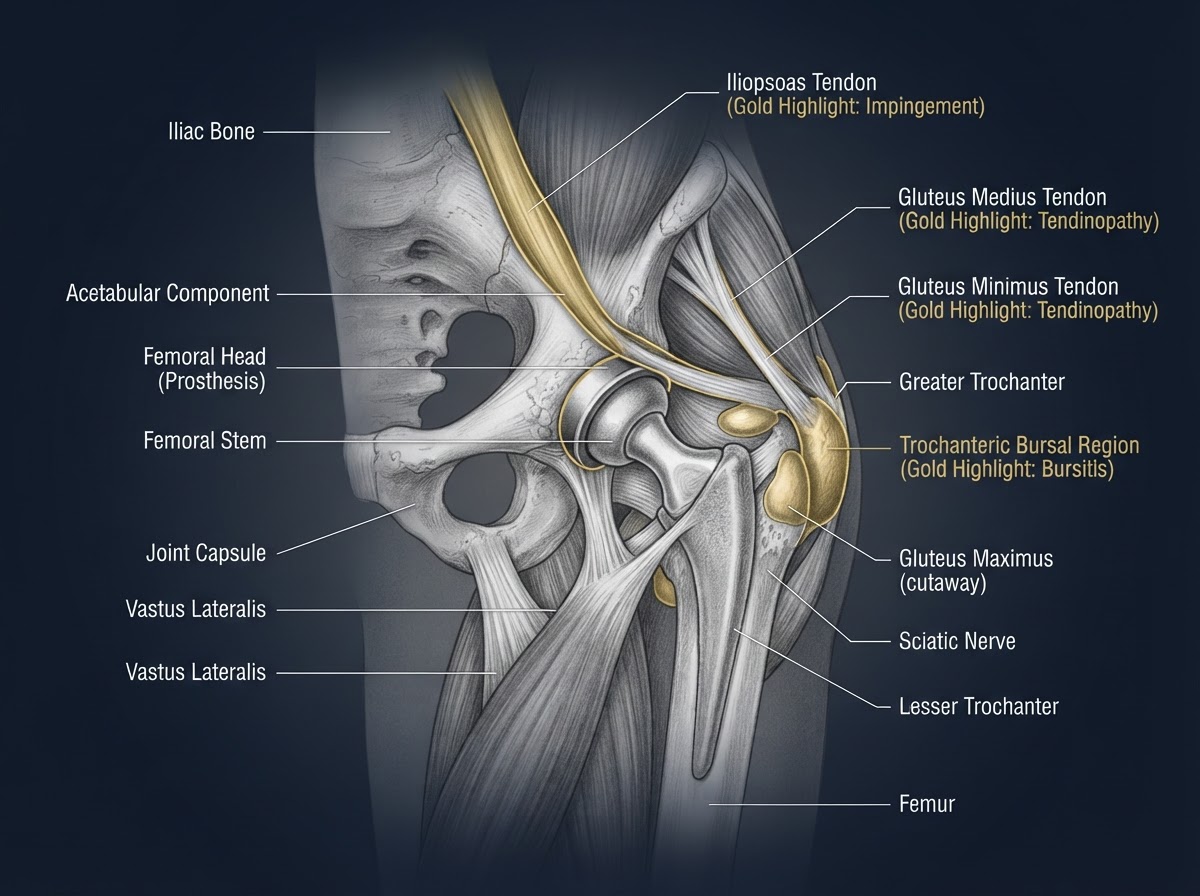
Anterior hip and groin pain is one of the most common pain patterns after replacement and has several distinct causes. Iliopsoas tendinopathy and iliopsoas impingement against the acetabular component are increasingly recognised contributors: the tendon crosses in front of the anterior cup, and a prominent or retroverted cup, screw prominence or simply the geometry of the reconstruction can irritate it. The typical clinical pattern is pain during active hip flexion — lifting the leg, climbing stairs, getting in and out of a car, or resisted hip flexion against the examiner's hand. Other causes of groin pain include component loosening, periprosthetic infection and other intra-articular or extra-articular pathology. Clinical examination and imaging help distinguish iliopsoas pathology from implant-related causes: an image-guided diagnostic injection around the tendon that produces a meaningful temporary response supports the diagnosis. Treatment ranges from conservative management and targeted physiotherapy to, in carefully selected cases with confirmed cup prominence, tendon release or component revision. It is important not to assume that all groin pain after hip replacement is iliopsoas impingement — infection and loosening must always be considered, and the diagnosis is confirmed by combining the clinical pattern, imaging and the response to targeted intervention.
Section 5: Lateral hip pain: gluteal tendons and greater trochanteric pain
Lateral hip pain over the greater trochanter is common and is often lumped under the label 'trochanteric bursitis', but the modern term 'greater trochanteric pain syndrome' better reflects that the pain usually arises from the gluteus medius and minimus tendons and their attachment, with trochanteric bursitis as a possible component rather than the whole story. Typical features include lateral hip pain, tenderness over the greater trochanter, pain when lying on the affected side, pain during single-leg stance or climbing stairs, and sometimes a limp or pelvic instability from abductor weakness. Causes include gluteus medius and minimus tendinopathy, partial or complete abductor tendon tears, altered biomechanics, and soft-tissue damage or repair-related problems from the surgical approach. Examination, ultrasound and MRI with metal-artifact reduction help characterise the tendons and rule out a significant tear. Treatment options include targeted physiotherapy, load modification, progressive abductor strengthening, selected image-guided interventions around the extra-articular soft tissues, and surgical repair in selected cases of significant abductor tears. Not every lateral pain syndrome requires surgery — many patients improve substantially with a structured rehabilitation programme — and injections around the trochanteric region must be distinguished from injections into the prosthetic joint itself (see Section 16).
Section 6: Posterior hip and buttock pain
Posterior hip and buttock pain after replacement is frequently not coming from the hip at all. Lumbar radiculopathy, lumbar spinal stenosis, facet-mediated pain, sacroiliac region pain and deep gluteal pathology can all produce pain in the buttock that the patient attributes to the hip. This is why a lumbar spine assessment is essential when the hip implant imaging is satisfactory but the pain persists. Clues suggesting a spinal origin include pain radiating below the knee, tingling or numbness, neurological symptoms, symptoms that change with spinal position or sitting, and back pain accompanying the hip-region discomfort. Pain location alone is insufficient to distinguish these conditions: a patient with a well-fixed, non-infected hip can have buttock and thigh pain referred entirely from the lumbar spine, and treating the hip will not help. A careful neurological and lumbar examination, alongside the hip work-up, prevents the common error of investigating — or even revising — a hip that is not the source of the pain.
Section 7: Thigh pain after hip replacement
Thigh pain raises the question of the femoral component. Early, mild, aching thigh discomfort can occur in the first months after an uncemented stem as the bone adapts to the implant and usually settles. Persistent or progressive thigh pain, however, demands investigation: it may reflect femoral component-related pain, aseptic loosening, a stress reaction, a periprosthetic fracture, bone remodelling or referred lumbar pain, and the cause depends on stem design and fixation, bone quality and the clinical timeline. It is important to distinguish early postoperative thigh discomfort — often self-limiting — from persistent or worsening pain, which should not be dismissed. The most common diagnostic error is to label every thigh pain as femoral stem loosening: the diagnosis requires radiographic and sometimes advanced imaging correlation, not symptoms alone. Comparison with previous radiographs is essential, because a stable stem that has not changed position over years is unlikely to be the source of new thigh pain even if it looks slightly different from the contralateral side.
Section 8: Aseptic loosening of the hip prosthesis
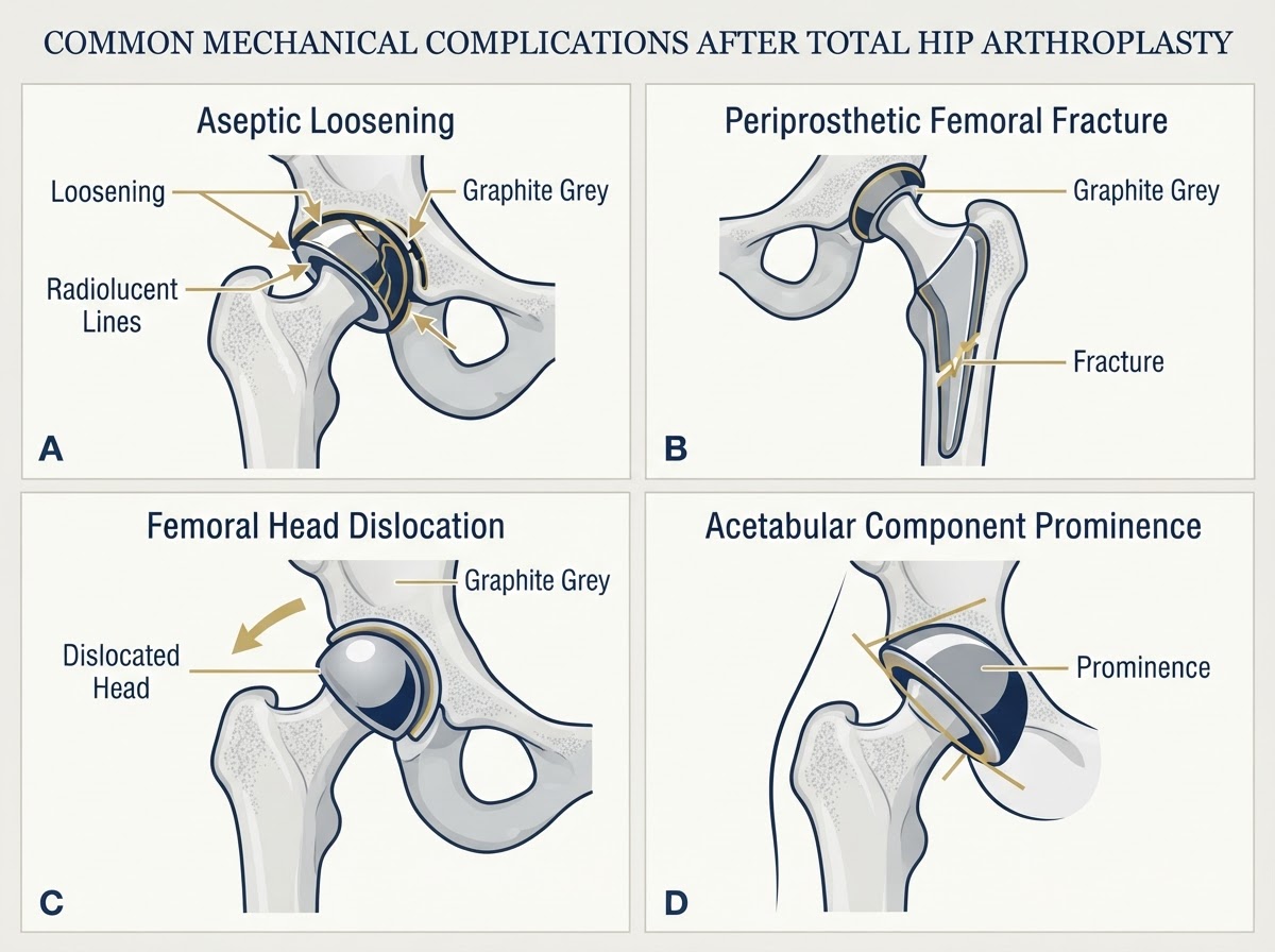
Aseptic loosening means the mechanical failure of implant fixation at the bone–implant interface, without infection. It is distinct from infection and the two must not be confused, because their management is entirely different. Loosening can affect the acetabular component, the femoral component, or both, and is often associated with osteolysis — progressive bone resorption around the implant driven by wear-particle debris. Radiographically, loosening may appear as progressive radiolucent lines around the component, component migration or tilt, or changing position on serial radiographs. Typical clinical features include progressive pain, pain that is worse on weight-bearing, start-up pain in some patients (pain on the first steps after rising), and a deterioration in walking tolerance. Crucially, radiographic findings must be interpreted alongside symptoms, previous images and implant characteristics: a small radiolucent line that has been stable for years is not the same as one that is clearly progressing. When conventional radiographs are inconclusive, further investigations such as CT or, in selected cases, nuclear imaging may help. Loosening is not diagnosed from pain alone — a patient with pain and normal, stable imaging has another cause that must be found. How the implant, its fixation and bearing surfaces behave over time is relevant context, but it does not by itself establish that a specific painful hip is loose.
Section 9: Periprosthetic joint infection
Periprosthetic joint infection is one of the most important diagnoses not to miss, and it can present at almost any time: early after surgery, during the first postoperative months, or years after implantation. Symptoms may include increasing pain, persistent wound drainage, redness and warmth, swelling, and progressive loss of function; fever may be present but is frequently absent, and low-grade infection can present with relatively subtle symptoms that mimic aseptic loosening. This is why infection must be considered before assuming aseptic loosening or proceeding with revision surgery. The diagnostic work-up combines clinical examination with inflammatory markers (C-reactive protein and erythrocyte sedimentation rate), joint aspiration with synovial white blood cell count and differential neutrophil percentage, and microbiological cultures; additional validated biomarkers may be used according to the clinical context. Modern diagnostic criteria — the 2018 International Consensus Meeting (ICM/MSIS) definition and the European Bone and Joint Infection Society (EBJIS) definition — provide structured, evidence-based frameworks, but laboratory thresholds depend on clinical context, timing since surgery and the specific criteria applied; they must not be interpreted in isolation. A normal CRP does not exclude all infections, and antibiotics before sampling can affect microbiological diagnosis. Because the management of infection (often a combination of antibiotic and surgical strategies selected according to infection type, timing, organism and implant stability) is so different from the management of aseptic loosening, excluding infection is a prerequisite, not an afterthought.
Section 10: Periprosthetic fracture
A periprosthetic fracture is a break in the bone around the implant — around the femoral stem or, less commonly, the acetabular component. These fractures can occur after a fall or trauma, but occasionally they occur without major trauma, particularly in patients with reduced bone quality or osteoporosis. The typical presentation is sudden pain, an inability or difficulty bearing weight, and sometimes deformity or shortening of the limb, with functional deterioration. Radiographs are the first-line investigation; CT may be necessary for complex patterns or planning. The Vancouver classification is the standard system for periprosthetic femoral fractures: it groups fractures by their location relative to the stem, the stability of the stem, and the quality of the surrounding bone stock, and it directly guides management. Treatment depends on the fracture pattern, stem stability, bone stock and the patient's overall condition, and ranges from protected weight-bearing to internal fixation or stem revision. A suspected periprosthetic fracture requires urgent assessment — patients should not attempt to walk through sudden, severe pain around the implant.
Section 11: Instability and hip dislocation
Recurrent instability and acute dislocation are important causes of pain and functional loss after hip replacement. Dislocation risk depends on component orientation, soft-tissue tension, abductor function, spinopelvic mechanics, previous spinal fusion (which alters pelvic mobility) and patient-specific factors. An acute dislocation typically presents with sudden severe pain, an inability to move or bear weight, and an abnormal limb position; recurrent episodes of instability may present with repeated giving-way sensations. Suspected dislocation requires emergency assessment and imaging — patients should not attempt to reduce the hip themselves. Management depends on the cause: a single dislocation may be managed with closed reduction and a period of protected mobility, while recurrent instability requires assessment of component position, soft-tissue tension and spinopelvic factors, and may require revision surgery to address the underlying mechanical problem. Instability is not something to 'live with': each dislocation episode carries risk, and recurrent instability deserves a structured assessment.
Section 12: Other causes of pain after hip replacement
Several less common but recognised causes complete the differential diagnosis. Heterotopic ossification — bone forming in the soft tissues around the hip — can cause stiffness and discomfort after surgery. Adverse local tissue reactions, relevant to certain implant types and bearing surfaces, can produce pain, effusion and soft-tissue damage; trunnion-related problems and corrosion at the head–neck junction are recognised in specific contexts. Neuropathic pain from nerve injury or irritation, and perceived or real leg-length discrepancy, can also drive persistent symptoms. Pain may even originate outside the operated hip entirely: contralateral hip osteoarthritis, knee pathology, or vascular and other non-orthopaedic causes can be misattributed to the replacement. The aim is not to overemphasise rare complications but to recognise when specialised implant investigations — advanced imaging, metal-ion testing where appropriate, or specialist revision assessment — are necessary. A careful history and examination usually prevent the common error of attributing all postoperative pain to the prosthesis.
Section 13: The clinical assessment: how the diagnosis begins
A systematic clinical evaluation is the foundation of the diagnosis, and it begins before any scan. The medical history captures the date of surgery, the surgical approach if known, the implant type if known, whether symptoms improved initially, the onset and progression of pain, the pain location, the relationship of pain to walking, loading and movement, any previous infections or revision surgery, and associated spine or joint conditions. The physical examination assesses gait, leg length, hip range of motion, resisted hip flexion (to screen for iliopsoas pain), abductor strength (to screen for abductor dysfunction), trochanteric tenderness, a neurological assessment, a lumbar spine assessment, and signs of instability where appropriate. Examination findings guide the choice of investigations: a patient with a normal gait, full painless range and trochanteric tenderness points towards a soft-tissue cause, while a patient with a painful antalgic gait and pain on weight-bearing points towards a component or structural problem. No single test replaces this clinical assessment.
Section 14: Imaging: which tests are useful?
Imaging is selected according to the clinical suspicion, not ordered as a blanket panel. Plain radiographs — an AP pelvis and appropriate lateral hip views — remain the first and often most useful investigation: they show component position, radiolucent lines, migration, osteolysis and fracture, and they are most informative when compared with previous images. CT adds detail on component position, acetabular version and prominence, bone loss, fracture and complex revision planning, and is particularly useful when radiographs are inconclusive. Ultrasound is excellent for the extra-articular soft tissues — the iliopsoas tendon, gluteal tendons, trochanteric region and soft-tissue fluid collections — and can guide aspiration or injection. MRI with metal-artifact reduction sequences characterises abductor tendon pathology, soft-tissue abnormalities, adverse local tissue reactions and selected infection-related findings that radiographs and CT cannot. Nuclear imaging (bone scintigraphy, labelled leukocyte imaging and related techniques) has selected roles when the picture is unclear, but findings must be interpreted carefully and in context. Not every patient needs every test: the right imaging is the one that answers the specific clinical question raised by the history and examination.
Section 15: Blood tests and joint aspiration
Blood tests and joint aspiration are central to the infection work-up. C-reactive protein (CRP) and erythrocyte sedimentation rate (ESR) are the standard inflammatory markers used to screen for infection; a complete blood count may be indicated. When inflammatory markers are raised or the clinical suspicion is high, joint aspiration provides synovial fluid for a white blood cell count, differential neutrophil percentage, cultures and additional tests according to the clinical suspicion. Two practical points matter. First, antibiotics given before sampling can suppress microbiological culture and make diagnosis harder — this is a reason to obtain samples before starting empirical antibiotics, but patients should not stop prescribed medication on their own and should follow their surgical team's instructions. Second, aspiration is an invasive procedure that must be selected and performed by an appropriately qualified clinician, usually under image guidance, because an inadequately performed aspiration risks introducing infection into a previously uninfected joint. Laboratory results are interpreted against validated diagnostic criteria and the clinical context, never in isolation.
Section 16: Diagnostic infiltrations and targeted injections
Image-guided injections have a selected diagnostic role. An injection around the iliopsoas sheath or bursa, a peritrochanteric injection, or a selected periarticular injection can help localise a pain generator: a meaningful temporary response supports a specific soft-tissue diagnosis and can guide further management. Two principles are essential. First, a temporary response localises a pain generator but does not prove the entire diagnosis — the whole clinical picture still matters. Second, and critically, injections should not be performed routinely into a painful prosthetic joint without first considering and appropriately excluding periprosthetic infection, because injecting steroids into an infected joint can seriously worsen the situation. It is important to distinguish injections around extra-articular structures (tendon sheaths, bursae) from injections into the prosthetic joint itself. PRP, corticosteroid and hyaluronic acid injections into a prosthetic joint are not established treatments for the painful hip replacement and should not be presented as such; their role, if any, is limited and unproven in this specific context.
Section 17: Treatment: why it must match the cause
Treatment is organised by diagnosis, because the right treatment for one cause is the wrong treatment for another. Extra-articular soft-tissue causes (abductor tendinopathy, trochanteric pain) are managed with activity modification, targeted rehabilitation, progressive strengthening, selected image-guided interventions around the soft tissues, and surgical repair only in selected significant tears. Iliopsoas impingement is managed with conservative measures and a diagnostic injection first, with evaluation of cup prominence and position, and tendon release or component revision reserved for carefully selected cases where anatomy and clinical findings support it. Aseptic loosening is managed with observation in selected asymptomatic or minimally symptomatic situations, further investigation when the picture is unclear, and revision surgery when clinically indicated. Periprosthetic infection requires specialist infection assessment, microbiological diagnosis and multidisciplinary management, with antibiotic and surgical strategies selected according to infection type, timing, organism and implant stability. Periprosthetic fracture is managed according to its classification, component stability and bone stock. Instability is managed by assessing component position, soft tissues and spinopelvic factors, with individualised treatment. The unifying principle is that revision surgery should not be recommended until the underlying cause has been adequately investigated — revising a well-fixed, non-infected hip for a soft-tissue or spinal problem will not relieve the pain.
Section 18: When is revision hip replacement necessary?
Revision hip replacement is a major undertaking and is considered for specific, confirmed indications: symptomatic aseptic loosening with progressive radiographic change; periprosthetic joint infection; recurrent instability that has not responded to non-operative management; significant component malposition with symptoms; periprosthetic fracture; selected adverse local tissue reactions; and other established mechanical failures. Crucially, not every painful hip replacement requires revision. A patient with abductor tendinopathy, iliopsoas impingement or lumbar referred pain may have a perfectly well-fixed, non-infected prosthesis, and the correct treatment targets the actual pain generator, not the implant. The distinction between treating a soft-tissue or spinal problem and replacing a well-fixed prosthesis is one of the most important judgements in revision arthroplasty, and it is exactly why a structured diagnostic work-up precedes any decision to revise. When revision is indicated, it is planned around the specific cause — loosening, infection, instability or fracture each demand a different strategy — and patients can read more about the procedure itself in our dedicated revision hip replacement article. For hips that are sound, the goal is a return to activity, and our guide to life after hip replacement covers walking, exercise and sport in detail.
Section 19: Warning signs: when to seek urgent assessment
Most persistent pain after hip replacement is not an emergency, but certain symptoms warrant urgent assessment: sudden severe hip pain; a new inability to bear weight; a suspected dislocation (sudden pain with an abnormal limb position); significant trauma such as a fall; fever with a painful or swollen hip; wound drainage or increasing redness; sudden calf swelling or pain; and chest pain or breathlessness. The last two are especially important: chest pain, sudden breathlessness or other symptoms suggestive of a thromboembolic complication require emergency care, and in Spain the emergency number is 112. The aim is not to make every patient anxious — most postoperative discomfort is benign and improves — but to ensure that the small number of genuinely urgent complications are identified and treated promptly rather than 'waited out'. For ordinary, gradually improving symptoms, a scheduled specialist review is appropriate; for sudden, severe or systemic symptoms, urgent assessment is the right choice.
Section 20: A practical diagnostic pathway
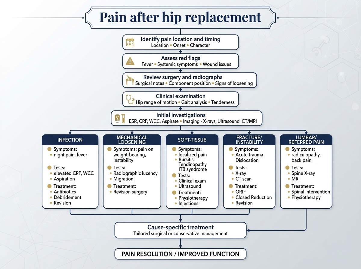
The diagnostic process can be summarised as a structured pathway that moves from the clinical picture to cause-specific treatment: 1. Pain after hip replacement is reported. 2. Identify the pain location and timing — groin, lateral, posterior, thigh; early, persistent, new, or post-trauma. 3. Assess red flags and urgent complications — dislocation, fracture, infection, thromboembolism. 4. Review the surgery, implant and previous radiographs. 5. Perform a focused clinical examination — gait, range, abductors, iliopsoas, lumbar spine, neurology. 6. Order initial investigations — radiographs and inflammatory markers where indicated. 7. Pursue targeted investigations according to the suspected cause: - Infection pathway: inflammatory markers, aspiration, cultures. - Mechanical loosening pathway: serial radiographs, CT, comparison with prior images. - Soft-tissue pathway: ultrasound, MRI with metal-artifact reduction, diagnostic injection. - Fracture/instability pathway: radiographs, CT, urgent assessment. - Lumbar/referred pain pathway: lumbar examination and imaging. 8. Apply cause-specific treatment matched to the confirmed diagnosis. This pathway is educational and supports, but never replaces, individual clinical assessment by a qualified orthopaedic surgeon.
Section 21: Frequently asked questions
Concise answers to the questions patients ask most often about pain after hip replacement: 1. Is it normal to have pain years after hip replacement? Not 'normal', but not uncommon — and it does not automatically mean the implant has failed. Persistent or new pain years later deserves investigation to identify the cause. 2. Why does my groin hurt after hip replacement? Groin pain has several causes, including iliopsoas tendinopathy or impingement, infection and loosening; the clinical pattern and imaging help distinguish them. 3. Why does the outside of my hip hurt? Lateral pain often comes from the gluteal tendons or greater trochanteric pain syndrome, not the implant itself; abductor weakness and lying on the side are typical clues. 4. Can the iliopsoas tendon cause pain after hip replacement? Yes — irritation where the tendon crosses the front of the acetabular component can cause pain on active hip flexion, stairs and getting in and out of a car. 5. Can back problems cause pain around a hip replacement? Yes — lumbar radiculopathy and spinal stenosis can refer pain to the buttock and thigh even when the hip implant is fine. 6. How do I know if my hip prosthesis is loose? Loosening is suggested by progressive weight-bearing pain and start-up pain and is confirmed by progressive radiographic change on serial X-rays, not by symptoms alone. 7. Can a hip replacement become infected years later? Yes — low-grade or haematogenous infection can appear years after surgery, sometimes with subtle symptoms; inflammatory markers and aspiration help diagnose it. 8. What blood tests are used to investigate infection? CRP and ESR are the standard screening markers; if raised or suspicious, joint aspiration with cell count and cultures follows. 9. Can an X-ray detect a loose hip replacement? X-rays often show loosening through radiolucent lines or migration, especially when compared with previous images; CT may be needed when X-rays are inconclusive. 10. When is CT or MRI needed? CT details component position, bone loss and fracture; MRI with metal-artifact reduction assesses abductor tendons and soft tissues; both are selected by the clinical question. 11. Can an ultrasound help diagnose pain after hip replacement? Yes — ultrasound is excellent for extra-articular soft tissues (iliopsoas, gluteal tendons, fluid collections) and can guide aspiration or injection. 12. Does every painful hip replacement require revision surgery? No — many painful hips are caused by soft-tissue, iliopsoas or lumbar problems and improve without revision; revision is reserved for confirmed indications. 13. Can I receive an injection around a hip replacement? Injections around extra-articular soft tissues can help diagnostically; injections into the prosthetic joint are not routine and must exclude infection first. 14. What is a periprosthetic fracture? A break in the bone around the implant, usually after a fall, classified by the Vancouver system and managed according to fracture pattern and stem stability. 15. What should I do if I suddenly cannot bear weight? Seek urgent assessment — sudden inability to bear weight may indicate dislocation, fracture or infection and should not be 'waited out'. 16. When should I seek a second opinion? When the cause of persistent pain is unclear, when revision has been recommended without a clear diagnosis, or when you want an independent assessment of your imaging and options. 17. Can a painful hip replacement be treated without replacing the implant? Often, yes — targeted physiotherapy, soft-tissue treatment, lumbar care or image-guided interventions can relieve pain when the implant itself is sound.
Identify the Cause Before Choosing the Treatment
Persistent or new pain after a hip replacement is unsettling, but it is not a verdict on the implant. The most important message of this guide is that pain after hip replacement requires a structured diagnostic approach: the location and timing of pain provide useful clues, implant-related and extra-articular causes must be distinguished, infection must be considered before assuming aseptic loosening, and imaging and laboratory tests should be selected according to the clinical suspicion rather than ordered as a blanket panel. Not every painful prosthesis needs revision surgery — many patients have a well-fixed, non-infected hip and a treatable soft-tissue or spinal cause — and accurate diagnosis should always precede treatment. If your pain has not been adequately explained, or if revision has been suggested without a clear diagnosis, a specialist assessment or an independent second opinion can help establish the real cause and the right path forward. The goal is not to reassure blindly, but to diagnose precisely and treat the actual problem.
References & scientific evidence
- Parvizi J, Tan TL, Goswami K, Higuera C, Della Valle C, Chen AF, Shohat N. The 2018 Definition of Periprosthetic Hip and Knee Infection: An Evidence-Based and Validated Criteria for Diagnosing Periprosthetic Joint Infection.. The Journal of Bone and Joint Surgery, 2018.Clinical guidelineDOI: 10.2106/JBJS.17.01432PMID: 29551303View source
- Perez-Prieto D, Portillo ME, Puig-Verdié L, Alier A, Gamba C, Horcajada JP, Soriano A, Font-Vaquer A, Mensa J, Martínez-Pastor JC, Bori G, Bausili C, Ballesteros M, Trampuz A, Puig L. The EBJIS definition of periprosthetic joint infection.. The Bone & Joint Journal, 2021.Clinical guidelineDOI: 10.1302/0301-620X.103B1.BJJ-2020-1381.R1PMID: 33380199View source
- American Academy of Orthopaedic Surgeons. Diagnosis and Prevention of Periprosthetic Joint Infections — Clinical Practice Guideline.. American Academy of Orthopaedic Surgeons (AAOS), 2019.Clinical guidelineView source
- Duncan CP, Masri BA. Fractures of the femur after hip replacement: the Vancouver classification.. Instructional Course Lectures, 1995.Observational studyPMID: 15057082View source
- Kunutsor SK, Barrett MC, Beswick AD et al.. Risk factors for dislocation after primary total hip replacement: meta-analysis of 125 studies involving approximately five million hip replacements.. The Lancet Rheumatology, 2019.Meta-analysisDOI: 10.1016/S2665-9913(19)30045-1View source
- National Institute for Health and Care Excellence. Joint replacement (primary): hip, knee and shoulder. NICE guideline NG157.. National Institute for Health and Care Excellence (NICE), 2020.Clinical guidelineView source
- Loppini M, Falez F, Della Rocca A, Di Martino A, Cavallo M, Grappiolo G, Perna A, Traina F. Incidence and Risk Factors of Iliopsoas Tendinopathy After Total Hip Arthroplasty.. The Journal of Arthroplasty, 2023.Observational studyDOI: 10.1016/j.arth.2023.01.014View source
- Fearon AM, Cook JL, Scarvell JM, Neeman T, Cormick W, Smith PN. Greater trochanteric pain syndrome: defining the clinical syndrome.. British Journal of Sports Medicine, 2013.Observational studyDOI: 10.1136/bjsports-2012-091570View source
- Fritz J, Lurie B, Miller TT, Potter HG. Imaging of hip arthroplasty-related complications: multimodality approach with an emphasis on metal artifact reduction MRI.. Seminars in Musculoskeletal Radiology, 2015.Observational studyDOI: 10.1055/s-0035-1547497View source
- Cyteval C, Bourdon A. Imaging of the painful hip after hip arthroplasty.. Diagnostic and Interventional Imaging, 2016.Observational studyDOI: 10.1016/j.diii.2016.05.013View source
- Garbuz DS, Tanzer M, Greidanus NV, Masri BA, Duncan CP. The John Charnley Award: Muscle damage and femoral fracture risk with the direct anterior approach to the hip.. Clinical Orthopaedics and Related Research, 2016.Observational studyDOI: 10.1007/s11999-016-4851-0View source
- Cooper HJ, Della Valle CJ. Massive acetabular erosion and cup migration after hip resurfacing: a case report.. HSS Journal, 2010.Observational studyDOI: 10.1007/s11420-010-9169-7View source
- Wainwright TW et al.. Consensus statement for perioperative care in total hip replacement and total knee replacement surgery: Enhanced Recovery After Surgery (ERAS®) Society recommendations.. Acta Orthopaedica, 2019.Clinical guidelineDOI: 10.1080/17453674.2019.1683790View source
- National Joint Registry for England, Wales, Northern Ireland and the Isle of Man. National Joint Registry 21st Annual Report — Hip and knee arthroplasty outcomes.. National Joint Registry (NJR), 2024.Observational studyView source
- European Bone and Joint Infection Society (EBJIS). EBJIS — European Bone & Joint Infection Society: periprosthetic joint infection resources.. European Bone and Joint Infection Society (EBJIS), 2024.Clinical guidelineView source
Frequently asked questions

Dr. Pablo Banchero Lasalvia
Orthopaedic Surgeon · Traumatology Team
- Author:
- Dr. Pablo Banchero Lasalvia
- Medical reviewer:
- Dr. Pablo Banchero Lasalvia
- Published:
- 4 November 2026
- Medical review date:
- 4 November 2026
- ·
- 20 min read
A Specialist Assessment Can Identify the Real Cause of Your Pain
A specialist assessment can help identify whether symptoms are related to the implant, surrounding soft tissues or another source. Discuss your symptoms and imaging with Dr Pablo Banchero.
Official number: +34 602 99 22 22